Sunday Poster Session
Category: Small Intestine
P1297 - Something "Fishy" in the Small Bowel: A Rare Case of Presentation of Meckel's Diverticulum
Sunday, October 22, 2023
3:30 PM - 7:00 PM PT
Location: Exhibit Hall

Has Audio

Annie Shergill, MD
Larkin Community Hospital
Miami, FL
Presenting Author(s)
Annie Shergill, MD1, Luis Geada, MD2, Luis Nasiff, MD3, Micaella Kantor, MD3
1Larkin Community Hospital, Miami, FL; 2Baptist Health Hospital, Miami, FL; 3Larkin Community Hospital, Palm Springs Campus, Miami, FL
Introduction: Meckel's diverticulum (MD) is a true diverticulum resulting from small bowel (SB) dysgenesis (incomplete obliteration of vitelline duct). Symptomatic MD is most commonly seen in children but it may remain completely asymptomatic & present insidiously in adult life with an associated complication such as SB obstruction. We present a unique case of presentation of MD in an adult with foreign body induced perforation of MD.
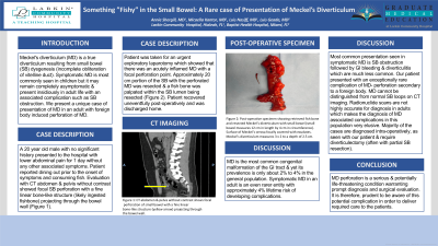
Case Description/Methods: A 20 year old male with no significant history presented to the hospital with lower abdominal pain for 1 day without any other associated symptoms. Patient reported dining out prior to the onset of symptoms and consuming fish. Evaluation with CT abdomen & pelvis without contrast showed focal SB perforation with a fine linear bone-like structure (likely ingested fishbone) projecting through the bowel wall (Figure 1B). Patient was taken for an urgent exploratory laparotomy which showed that there was an acutely inflamed MD with a focal perforation point. Approximately 20 cm portion of the SB with the perforated MD was resected & a fish bone was palpated within the SB lumen being resected (Figure 1A). Patient recovered uneventfully post-operatively and was discharged home.
Discussion: MD is the most common congenital malformation of the GI tract & yet its prevalence is only about 2% to 4% in the general population. Symptomatic MD in an adult is an even rarer entity with approximately 4% lifetime risk of developing complications. Most common presentation seen in symptomatic MD is SB obstruction followed by GI bleeding & diverticulitis which are much less common. Our patient presented with an exceptionally rare complication of MD- perforation secondary to a foreign body. MD cannot be distinguished from normal SB loops on CT imaging. Radionuclide scans are not highly accurate for diagnosis in adults which makes the diagnosis of MD associated complications in this population very elusive. Majority of the cases are diagnosed intra-operatively, as seen with our patient & require diverticulectomy (often with partial SB resection). MD perforation is a serious & potentially life-threatening condition warranting prompt diagnosis and surgical evaluation. It is therefore, prudent to be aware of this potential complication in order to deliver required care to the patients.

Disclosures:
Annie Shergill, MD1, Luis Geada, MD2, Luis Nasiff, MD3, Micaella Kantor, MD3. P1297 - Something "Fishy" in the Small Bowel: A Rare Case of Presentation of Meckel's Diverticulum, ACG 2023 Annual Scientific Meeting Abstracts. Vancouver, BC, Canada: American College of Gastroenterology.
1Larkin Community Hospital, Miami, FL; 2Baptist Health Hospital, Miami, FL; 3Larkin Community Hospital, Palm Springs Campus, Miami, FL
Introduction: Meckel's diverticulum (MD) is a true diverticulum resulting from small bowel (SB) dysgenesis (incomplete obliteration of vitelline duct). Symptomatic MD is most commonly seen in children but it may remain completely asymptomatic & present insidiously in adult life with an associated complication such as SB obstruction. We present a unique case of presentation of MD in an adult with foreign body induced perforation of MD.
Case Description/Methods: A 20 year old male with no significant history presented to the hospital with lower abdominal pain for 1 day without any other associated symptoms. Patient reported dining out prior to the onset of symptoms and consuming fish. Evaluation with CT abdomen & pelvis without contrast showed focal SB perforation with a fine linear bone-like structure (likely ingested fishbone) projecting through the bowel wall (Figure 1B). Patient was taken for an urgent exploratory laparotomy which showed that there was an acutely inflamed MD with a focal perforation point. Approximately 20 cm portion of the SB with the perforated MD was resected & a fish bone was palpated within the SB lumen being resected (Figure 1A). Patient recovered uneventfully post-operatively and was discharged home.
Discussion: MD is the most common congenital malformation of the GI tract & yet its prevalence is only about 2% to 4% in the general population. Symptomatic MD in an adult is an even rarer entity with approximately 4% lifetime risk of developing complications. Most common presentation seen in symptomatic MD is SB obstruction followed by GI bleeding & diverticulitis which are much less common. Our patient presented with an exceptionally rare complication of MD- perforation secondary to a foreign body. MD cannot be distinguished from normal SB loops on CT imaging. Radionuclide scans are not highly accurate for diagnosis in adults which makes the diagnosis of MD associated complications in this population very elusive. Majority of the cases are diagnosed intra-operatively, as seen with our patient & require diverticulectomy (often with partial SB resection). MD perforation is a serious & potentially life-threatening condition warranting prompt diagnosis and surgical evaluation. It is therefore, prudent to be aware of this potential complication in order to deliver required care to the patients.

Figure: Figure 1A: Post-operative specimen showing retrieved fish bone and resected Meckel’s diverticulum with small bowel (small bowel measures 12 cm in length by 4 cm in circumference). Surface of Meckel’s serosa focally covered with exudates. Meckel’s diverticulum measures 3 x 2 to a depth of 2.5 cm. 1B: CT abdomen & pelvis without contrast shows focal perforation of small bowel with a fine linear bone-like structure (yellow arrow) projecting through the bowel wall.
Disclosures:
Annie Shergill indicated no relevant financial relationships.
Luis Geada indicated no relevant financial relationships.
Luis Nasiff indicated no relevant financial relationships.
Micaella Kantor indicated no relevant financial relationships.
Annie Shergill, MD1, Luis Geada, MD2, Luis Nasiff, MD3, Micaella Kantor, MD3. P1297 - Something "Fishy" in the Small Bowel: A Rare Case of Presentation of Meckel's Diverticulum, ACG 2023 Annual Scientific Meeting Abstracts. Vancouver, BC, Canada: American College of Gastroenterology.
