Sunday Poster Session
Category: Small Intestine
P1301 - Rare Causes of Gastrointestinal Hemorrhage: Series of Adult Duodenal and Jejunal Gastric Heterotopia
Sunday, October 22, 2023
3:30 PM - 7:00 PM PT
Location: Exhibit Hall

Has Audio

Venkata Vinod Kumar Matli, MD
Christus Highland Medical Center
Shreveport, Louisiana
Presenting Author(s)
Venkata Vinod Kumar Matli, MD1, Andre Morgan, MD1, Dustin Hadley, PA-C2, Varsha Pujala, MD1, Kirtesh Patel, MD1, John Kirkikis, MD3
1Christus Highland Medical Center, Shreveport, LA; 2GastroIntestinal Specialist, Shreveport, LA; 3GastroIntestinal Specialists, Shreveport, LA
Introduction: Heterotopia refers to organ displacement to a non-physiological site. Gastric heterotopia (GH) is a rare cause of obscure gastrointestinal bleeding. GH of the small bowel is very rare, and the duodenum is more commonly involved than the jejunum. Here, we present five cases of GH involving the duodenum and jejunum, with presentations including gastrointestinal bleeding, symptomatic anemia, and no symptoms.
Case Description/Methods: A 63-year-old man presented to our clinic with gastrointestinal hemorrhage. Upper endoscopy, wireless capsule endoscopy, and colonoscopy found no obvious evidence of the source of bleeding, and the patient was ultimately diagnosed with jejunal GH on histopathological examination.
A 70-year-old woman presented with melena. Upper endoscopy revealed a fleshy polypoid mass in the duodenal bulb, which was histopathologically diagnosed as GH.
A 50-year-old woman presented with nausea, vomiting, and dysphagia. Upper endoscopy showed grade II esophagitis at the gastroesophageal junction, and a single, median, semi-pedunculated polyp in the duodenal bulb. Histopathology showed GH, but was negative for dysplasia or malignancy.
A 69-year-old woman had an incidental finding of duodenal GH when she was evaluated for a right lung mass with metastatic liver disease. Unfortunately, this patient was diagnosed with an aggressive small cell neuroendocrine tumor of pulmonary origin.
An 83-year-old woman with symptomatic anemia upper endoscopy revealed duodenal polyps, and histopathological diagnosis was consistent with benign GH.
Discussion: GH is one of the rarest causes of obscure gastrointestinal bleeding, and its exact mechanism remains unknown. Its clinical presentation is variable and gastrointestinal bleeding is rare. Diagnosis is based only on histopathological studies. Our case series emphasizes the need to include GH in the differential diagnosis when patients present with abdominal pain, gastrointestinal bleeding, or asymptomatic incidental endoscopic polypoid findings. This will ensure timely diagnosis and minimize complications and mortality. Management varies from clinical observation to resection of the lesions through surgery, depending on size, location, and severity of symptomatology.

Disclosures:
Venkata Vinod Kumar Matli, MD1, Andre Morgan, MD1, Dustin Hadley, PA-C2, Varsha Pujala, MD1, Kirtesh Patel, MD1, John Kirkikis, MD3. P1301 - Rare Causes of Gastrointestinal Hemorrhage: Series of Adult Duodenal and Jejunal Gastric Heterotopia, ACG 2023 Annual Scientific Meeting Abstracts. Vancouver, BC, Canada: American College of Gastroenterology.
1Christus Highland Medical Center, Shreveport, LA; 2GastroIntestinal Specialist, Shreveport, LA; 3GastroIntestinal Specialists, Shreveport, LA
Introduction: Heterotopia refers to organ displacement to a non-physiological site. Gastric heterotopia (GH) is a rare cause of obscure gastrointestinal bleeding. GH of the small bowel is very rare, and the duodenum is more commonly involved than the jejunum. Here, we present five cases of GH involving the duodenum and jejunum, with presentations including gastrointestinal bleeding, symptomatic anemia, and no symptoms.
Case Description/Methods: A 63-year-old man presented to our clinic with gastrointestinal hemorrhage. Upper endoscopy, wireless capsule endoscopy, and colonoscopy found no obvious evidence of the source of bleeding, and the patient was ultimately diagnosed with jejunal GH on histopathological examination.
A 70-year-old woman presented with melena. Upper endoscopy revealed a fleshy polypoid mass in the duodenal bulb, which was histopathologically diagnosed as GH.
A 50-year-old woman presented with nausea, vomiting, and dysphagia. Upper endoscopy showed grade II esophagitis at the gastroesophageal junction, and a single, median, semi-pedunculated polyp in the duodenal bulb. Histopathology showed GH, but was negative for dysplasia or malignancy.
A 69-year-old woman had an incidental finding of duodenal GH when she was evaluated for a right lung mass with metastatic liver disease. Unfortunately, this patient was diagnosed with an aggressive small cell neuroendocrine tumor of pulmonary origin.
An 83-year-old woman with symptomatic anemia upper endoscopy revealed duodenal polyps, and histopathological diagnosis was consistent with benign GH.
Discussion: GH is one of the rarest causes of obscure gastrointestinal bleeding, and its exact mechanism remains unknown. Its clinical presentation is variable and gastrointestinal bleeding is rare. Diagnosis is based only on histopathological studies. Our case series emphasizes the need to include GH in the differential diagnosis when patients present with abdominal pain, gastrointestinal bleeding, or asymptomatic incidental endoscopic polypoid findings. This will ensure timely diagnosis and minimize complications and mortality. Management varies from clinical observation to resection of the lesions through surgery, depending on size, location, and severity of symptomatology.

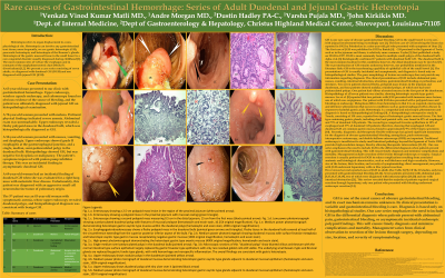
Figure: Figure 1a: Enteroscopy showing a 2.5-cm polypoid mass lesion in the region of the proximal jejunum (white pointed arrow) Figure 1b: Low power photomicrograph showing a pedunculated duodenal polyp with features of mucosal prolapse (hematoxylin and eosin stain, 10.25X original magnification) Figure 2a: Esophagogastroduodenoscopy shows a fleshy (pointed green arrows and triangles) polypoid mass in the duodenal bulb with. Fleshy tissue in the duodenal bulb that covered at least half of the circumference of bulb extending from the superior posterior inferior aspect of the bulb. Figure 2b: Medium power photomicrograph showing duodenal mucosa with surface foveolar metaplasia and heterotopic oxyntic type gastric mucosa recapitulating negative gastric mucosa (100 X original magnification, hematoxylin and eosin stain) Figure 3a: There was a single medium semi-pedunculated polyp in the duodenal bulb(Pointed arrow) Figure 3b: Microscopic sections of this "duodenal polyp" show blunted villous architecture with the normal intestinal type surface epithelium largely replaced by gastric foveolar- type epithelium with only are rare residual goblet cells still visible. The underlying small bowel crypts and Brunner glands are replaced by gastric fundic-type glands and with mild hemorrhage and nonspecific inflammation. The overall findings are consistent with gastric heterotopia. Figure 4a: Upper endoscopy shows (Pointed yellow arrow) nodular polyp in the duodenum. Figure 4b: Medium power photo micrograph of duodenal mucosa demonstrating heterotopic gastric oxyntic type glands adjacent to duodenal mucosal epithelium (Hematoxylin and Eosin stain, 100 X original magnification)
gastric heterotopia.jpg
gastric heterotopia.jpg
Disclosures:
Venkata Vinod Kumar Matli indicated no relevant financial relationships.
Andre Morgan indicated no relevant financial relationships.
Dustin Hadley indicated no relevant financial relationships.
Varsha Pujala indicated no relevant financial relationships.
Kirtesh Patel indicated no relevant financial relationships.
John Kirkikis indicated no relevant financial relationships.
Venkata Vinod Kumar Matli, MD1, Andre Morgan, MD1, Dustin Hadley, PA-C2, Varsha Pujala, MD1, Kirtesh Patel, MD1, John Kirkikis, MD3. P1301 - Rare Causes of Gastrointestinal Hemorrhage: Series of Adult Duodenal and Jejunal Gastric Heterotopia, ACG 2023 Annual Scientific Meeting Abstracts. Vancouver, BC, Canada: American College of Gastroenterology.
