Sunday Poster Session
Category: Small Intestine
P1305 - Chronic Perforated Appendicitis With Appendiceal Abscess Complicated by the Development of Vertebral Osteomyelitis
Sunday, October 22, 2023
3:30 PM - 7:00 PM PT
Location: Exhibit Hall

Has Audio

Samuel Addo, MD
UNC Health Blue Ridge
Morganton, NC
Presenting Author(s)
Samuel Addo, MD1, William Costello, DO2, Hayden Caraway, DO2, Dustin Kilpatrick, DO1, Rahul Sampath, MD1, Gerald Mank, MD1
1UNC Health Blue Ridge, Morganton, NC; 2UNC Health Blue Ridge Morganton, Morganton, NC
Introduction: Appendicitis is an inflammation of the appendix associated with luminal obstruction resulting in bacterial overgrowth and can lead to perforation. Chronic perforated appendicitis is a rare complication of appendicitis characterized by a delayed rupture of the appendix, leading to the formation of an appendiceal abscess. We present an elderly man admitted for vertebral osteomyelitis (VOM) with a CT abdomen incidentally demonstrating a large peri-appendiceal abscess and appendicitis.
Case Description/Methods: A 79-year-old male was referred to the emergency department by his primary care provider after a CT of the thoracic spine for chronic back pain revealed VOM. The patient reported that his symptoms started 4 months prior with intermittent low-grade fever and new onset diffuse lower abdominal pain for 3 weeks. The patient had a history of ischemic cardiomyopathy, obesity, and a bioprosthetic aortic valve. At baseline, he was ambulatory and independent with his daily activities.
On examination, the patient was hemodynamically stable, with diffuse lower abdominal tenderness without guarding and thoracic spine tenderness at the T7-T8 level. He had no neurological deficits, and the rest of his exam was unremarkable.
Laboratory investigations were concerning for elevated white blood cells, erythrocyte sedimentation rate, and C-reactive proteins.
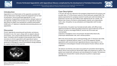
MRI of the thoracolumbar spine confirmed findings with T7-T8 discitis with VOM, no epidural or paraspinal abscess noted. CT scan of the abdomen showed appendicitis with a prominent peri-appendiceal abscess measuring 8.5 x 2 cm. Transthoracic echocardiogram showed normal ejection fraction without valvular vegetation.
The abscess was drained, and culture was positive for prevotella melaninogenica. Blood cultures were positive for Bacteroides fragilis and streptococcus species.The patient was treated with ceftriaxone and metronidazole for 6 weeks. Percutaneous drainage was removed after 4 weeks with plans to perform a laparoscopic appendectomy.
Discussion: Chronic appendicitis presenting with perforation and abscess formation is rare. Our case is unique with an atypical subacute presentation, and a polymicrobial bacteremia that seeded the spine. A scholarly search revealed no documented cases of VOM secondary to chronic appendicitis with appendiceal rupture. Complicated appendicitis with large abscess such as ours require placement of drainage for culture and antibiotic therapy followed by surgical resection of the appendix.

Disclosures:
Samuel Addo, MD1, William Costello, DO2, Hayden Caraway, DO2, Dustin Kilpatrick, DO1, Rahul Sampath, MD1, Gerald Mank, MD1. P1305 - Chronic Perforated Appendicitis With Appendiceal Abscess Complicated by the Development of Vertebral Osteomyelitis, ACG 2023 Annual Scientific Meeting Abstracts. Vancouver, BC, Canada: American College of Gastroenterology.
1UNC Health Blue Ridge, Morganton, NC; 2UNC Health Blue Ridge Morganton, Morganton, NC
Introduction: Appendicitis is an inflammation of the appendix associated with luminal obstruction resulting in bacterial overgrowth and can lead to perforation. Chronic perforated appendicitis is a rare complication of appendicitis characterized by a delayed rupture of the appendix, leading to the formation of an appendiceal abscess. We present an elderly man admitted for vertebral osteomyelitis (VOM) with a CT abdomen incidentally demonstrating a large peri-appendiceal abscess and appendicitis.
Case Description/Methods: A 79-year-old male was referred to the emergency department by his primary care provider after a CT of the thoracic spine for chronic back pain revealed VOM. The patient reported that his symptoms started 4 months prior with intermittent low-grade fever and new onset diffuse lower abdominal pain for 3 weeks. The patient had a history of ischemic cardiomyopathy, obesity, and a bioprosthetic aortic valve. At baseline, he was ambulatory and independent with his daily activities.
On examination, the patient was hemodynamically stable, with diffuse lower abdominal tenderness without guarding and thoracic spine tenderness at the T7-T8 level. He had no neurological deficits, and the rest of his exam was unremarkable.
Laboratory investigations were concerning for elevated white blood cells, erythrocyte sedimentation rate, and C-reactive proteins.
MRI of the thoracolumbar spine confirmed findings with T7-T8 discitis with VOM, no epidural or paraspinal abscess noted. CT scan of the abdomen showed appendicitis with a prominent peri-appendiceal abscess measuring 8.5 x 2 cm. Transthoracic echocardiogram showed normal ejection fraction without valvular vegetation.
The abscess was drained, and culture was positive for prevotella melaninogenica. Blood cultures were positive for Bacteroides fragilis and streptococcus species.The patient was treated with ceftriaxone and metronidazole for 6 weeks. Percutaneous drainage was removed after 4 weeks with plans to perform a laparoscopic appendectomy.
Discussion: Chronic appendicitis presenting with perforation and abscess formation is rare. Our case is unique with an atypical subacute presentation, and a polymicrobial bacteremia that seeded the spine. A scholarly search revealed no documented cases of VOM secondary to chronic appendicitis with appendiceal rupture. Complicated appendicitis with large abscess such as ours require placement of drainage for culture and antibiotic therapy followed by surgical resection of the appendix.

Figure: Figure 1. CT abdomen and pelvis findings consistent with appendicitis with prominent periappendiceal abscess measuring approximately 8.5 x 2.8 cm.
Disclosures:
Samuel Addo indicated no relevant financial relationships.
William Costello indicated no relevant financial relationships.
Hayden Caraway indicated no relevant financial relationships.
Dustin Kilpatrick indicated no relevant financial relationships.
Rahul Sampath indicated no relevant financial relationships.
Gerald Mank indicated no relevant financial relationships.
Samuel Addo, MD1, William Costello, DO2, Hayden Caraway, DO2, Dustin Kilpatrick, DO1, Rahul Sampath, MD1, Gerald Mank, MD1. P1305 - Chronic Perforated Appendicitis With Appendiceal Abscess Complicated by the Development of Vertebral Osteomyelitis, ACG 2023 Annual Scientific Meeting Abstracts. Vancouver, BC, Canada: American College of Gastroenterology.
