Sunday Poster Session
Category: Small Intestine
P1313 - Duodenal Pseudomelanosis in a Patient With Chronic Renal Disease
Sunday, October 22, 2023
3:30 PM - 7:00 PM PT
Location: Exhibit Hall


Saraswathi Lakkasani, MD
Saint Michael's Medical Center, New York Medical College
Glen Allen, VA
Presenting Author(s)
Saraswathi Lakkasani, MD1, Srinivas Pantula, MBBS2, Ummul Zohra Asfeen, DO3, Tahirunnisa Rida, BS4, Yashwitha Sai Pulakurthi, MBBS5, Yatinder Bains, MD6
1Saint Michael's Medical Center, New York Medical College, Newark, NJ; 2Saint Michael's Medical Center, Ontario, CA; 3Saint Michael's Medical Center, Newark, NJ; 4University of Texas at Dallas, Dallas, TX; 5New York Medical College-Saint Michael's Medical Center, Newark, NJ; 6Saint Michael's Medical Center, Newark, NJ
Introduction: Duodenal Pseudomelanosis (DP) is an asymptomatic and benign finding which is more commonly seen in middle aged to elderly females, characterized by the rare endoscopic appearance of discrete speckled black pigmentation of the duodenal mucosa. Certain medications such as ferrous sulfate, thiazides, vitamins, furosemide, digoxin and methyldopa have been linked to DP and some chronic medical conditions such as end stage renal disease, chronic hypertension, congestive heart failure, anemia have been reported to be associated with DP. The pathogenesis and its course are not yet clearly defined. Here we present a case of DP secondary to Chronic renal failure.
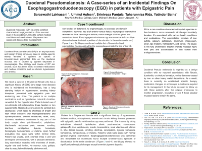
Case Description/Methods: A 56-year-old female with history of end stage renal disease on hemodialysis, hypertension, presented with Chronic epigastric pain and endoscopy was done. She is on Hydralazine, clonidine, Carvedilol for hypertension. Patient denied use of non-steroidal anti-inflammatory drugs, laxative or iron supplements. Denies any other complaints. Vital signs were within normal limits. Physical examination showed left arterio-venous fistula. Esophagogastroduodenoscopy was remarkable for multiple diminutive brownish-black speckles of discoloration in the entire examined duodenum. Biopsy confirmed multiple foci of brownish - black pigmentation within the macrophages inside the tips of the villi.
Discussion: Histology demonstrates a fine granular brown material inside the macrophage lysosomes in the lamina propria within the tips of the duodenal villi. While melanosis coli is secondary to the accumulation of lipofuscin in the macrophages of the colon, the pigment found in DP is composed mainly of iron sulfide, although varying amounts of sulfur, calcium, potassium, aluminum, magnesium, silver, lipo melanin, hemosiderin, ceroid and iron sulfide are also seen. Prussian blue and Fontana-Masson stains can be used to detect these pigments in macrophages. Tang et al. hypothesized mucosal injury caused by pills expose the gut macrophages to iron and other pigments and the duodenum is an absorptive organ for these pigments. Others have theorized that the sulfur component of various antihypertensives could lead to accumulation of iron sulfide, causing pigmentation. This condition is considered a benign condition that has not been related to fibrosis, duodenitis, or stricture formation unlike iron or other heavy metal deposition diseases. Hence, any specific therapy or change of medications and endoscopic surveillance have not been determined.

Disclosures:
Saraswathi Lakkasani, MD1, Srinivas Pantula, MBBS2, Ummul Zohra Asfeen, DO3, Tahirunnisa Rida, BS4, Yashwitha Sai Pulakurthi, MBBS5, Yatinder Bains, MD6. P1313 - Duodenal Pseudomelanosis in a Patient With Chronic Renal Disease, ACG 2023 Annual Scientific Meeting Abstracts. Vancouver, BC, Canada: American College of Gastroenterology.
1Saint Michael's Medical Center, New York Medical College, Newark, NJ; 2Saint Michael's Medical Center, Ontario, CA; 3Saint Michael's Medical Center, Newark, NJ; 4University of Texas at Dallas, Dallas, TX; 5New York Medical College-Saint Michael's Medical Center, Newark, NJ; 6Saint Michael's Medical Center, Newark, NJ
Introduction: Duodenal Pseudomelanosis (DP) is an asymptomatic and benign finding which is more commonly seen in middle aged to elderly females, characterized by the rare endoscopic appearance of discrete speckled black pigmentation of the duodenal mucosa. Certain medications such as ferrous sulfate, thiazides, vitamins, furosemide, digoxin and methyldopa have been linked to DP and some chronic medical conditions such as end stage renal disease, chronic hypertension, congestive heart failure, anemia have been reported to be associated with DP. The pathogenesis and its course are not yet clearly defined. Here we present a case of DP secondary to Chronic renal failure.
Case Description/Methods: A 56-year-old female with history of end stage renal disease on hemodialysis, hypertension, presented with Chronic epigastric pain and endoscopy was done. She is on Hydralazine, clonidine, Carvedilol for hypertension. Patient denied use of non-steroidal anti-inflammatory drugs, laxative or iron supplements. Denies any other complaints. Vital signs were within normal limits. Physical examination showed left arterio-venous fistula. Esophagogastroduodenoscopy was remarkable for multiple diminutive brownish-black speckles of discoloration in the entire examined duodenum. Biopsy confirmed multiple foci of brownish - black pigmentation within the macrophages inside the tips of the villi.
Discussion: Histology demonstrates a fine granular brown material inside the macrophage lysosomes in the lamina propria within the tips of the duodenal villi. While melanosis coli is secondary to the accumulation of lipofuscin in the macrophages of the colon, the pigment found in DP is composed mainly of iron sulfide, although varying amounts of sulfur, calcium, potassium, aluminum, magnesium, silver, lipo melanin, hemosiderin, ceroid and iron sulfide are also seen. Prussian blue and Fontana-Masson stains can be used to detect these pigments in macrophages. Tang et al. hypothesized mucosal injury caused by pills expose the gut macrophages to iron and other pigments and the duodenum is an absorptive organ for these pigments. Others have theorized that the sulfur component of various antihypertensives could lead to accumulation of iron sulfide, causing pigmentation. This condition is considered a benign condition that has not been related to fibrosis, duodenitis, or stricture formation unlike iron or other heavy metal deposition diseases. Hence, any specific therapy or change of medications and endoscopic surveillance have not been determined.

Figure: Endoscopic picture showing multiple dark pigmented spots in the duodenum
Disclosures:
Saraswathi Lakkasani indicated no relevant financial relationships.
Srinivas Pantula indicated no relevant financial relationships.
Ummul Zohra Asfeen indicated no relevant financial relationships.
Tahirunnisa Rida indicated no relevant financial relationships.
Yashwitha Sai Pulakurthi indicated no relevant financial relationships.
Yatinder Bains indicated no relevant financial relationships.
Saraswathi Lakkasani, MD1, Srinivas Pantula, MBBS2, Ummul Zohra Asfeen, DO3, Tahirunnisa Rida, BS4, Yashwitha Sai Pulakurthi, MBBS5, Yatinder Bains, MD6. P1313 - Duodenal Pseudomelanosis in a Patient With Chronic Renal Disease, ACG 2023 Annual Scientific Meeting Abstracts. Vancouver, BC, Canada: American College of Gastroenterology.
