Sunday Poster Session
Category: Small Intestine
P1322 - Retrograde Enteroscopy Revealing Terminal Ileum Adenocarcinoma
Sunday, October 22, 2023
3:30 PM - 7:00 PM PT
Location: Exhibit Hall

Has Audio

Anas Khouri, MD
University of South Alabama
Mobile, AL
Presenting Author(s)
Anas Khouri, MD1, Elizabeth G. Statham, BA2, Abrahim Hanjar, MD1, Phillip Henderson, DO1
1University of South Alabama, Mobile, AL; 2University of South Alabama College of Medicine, Mobile, AL
Introduction: Small bowel malignancy is rare, consisting of 1-3% of all primary GI malignancies. One third of those are further characterized as adenocarcinomas, with the terminal ileum being the least likely location to be affected (10% of small bowel adenocarcinomas). This report details a case of de novo terminal ileum adenocarcinoma, without underlying condition, in a patient presenting with iron deficiency anemia (IDA).
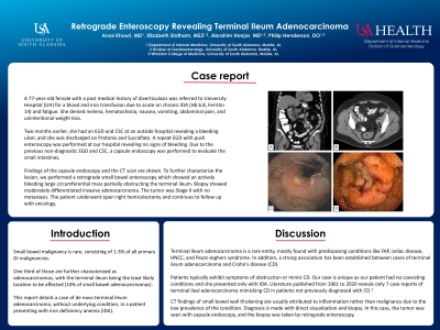
Case Description/Methods: A 77-year-old female with a past medical history of diverticulosis was referred to University Hospital (UH) for a blood and iron transfusion due to acute on chronic IDA (Hb 6.8, Ferritin 14) and fatigue. She denied melena, hematochezia, nausea, vomiting, abdominal pain, and unintentional weight loss. Two months earlier, she had an EGD and CSC at an outside hospital revealing a bleeding ulcer, and she was discharged on Protonix and Carafate. A repeat EGD with push enteroscopy was performed at our hospital revealing no signs of bleeding. Due to the previous non-diagnostic EGD and CSC, a capsule endoscopy was performed to evaluate the small intestines. Findings of the capsule endoscopy and the CT scan are shown below. To further characterize the lesion, we performed a retrograde small bowel enteroscopy which showed an actively bleeding large circumferential mass partially obstructing the terminal ileum. Biopsy showed moderately differentiated invasive adenocarcinoma. The tumor was Stage II with no metastasis. The patient underwent open right hemicolectomy and continues to follow up with oncology.
Discussion: Terminal ileum adenocarcinoma is a rare entity, mostly found with predisposing conditions like FAP, celiac disease, HNCC, and Peutz-Jeghers syndrome. In addition, a strong association has been established between cases of terminal ileum adenocarcinoma and Crohn’s disease (CD). Patients typically exhibit symptoms of obstruction or mimic CD. Our case is unique as our patient had no coexisting conditions and she presented only with IDA. Literature published from 1961 to 2020 reveals only 7 case reports of terminal ileal adenocarcinoma mimicking CD in patients not previously diagnosed with CD.1 CT findings of small bowel wall thickening are usually attributed to inflammation rather than malignancy due to the low prevalence of the condition. Diagnosis is made with direct visualization and biopsy. In this case, the tumor was seen with capsule endoscopy, and the biopsy was taken by retrograde enteroscopy.

Disclosures:
Anas Khouri, MD1, Elizabeth G. Statham, BA2, Abrahim Hanjar, MD1, Phillip Henderson, DO1. P1322 - Retrograde Enteroscopy Revealing Terminal Ileum Adenocarcinoma, ACG 2023 Annual Scientific Meeting Abstracts. Vancouver, BC, Canada: American College of Gastroenterology.
1University of South Alabama, Mobile, AL; 2University of South Alabama College of Medicine, Mobile, AL
Introduction: Small bowel malignancy is rare, consisting of 1-3% of all primary GI malignancies. One third of those are further characterized as adenocarcinomas, with the terminal ileum being the least likely location to be affected (10% of small bowel adenocarcinomas). This report details a case of de novo terminal ileum adenocarcinoma, without underlying condition, in a patient presenting with iron deficiency anemia (IDA).
Case Description/Methods: A 77-year-old female with a past medical history of diverticulosis was referred to University Hospital (UH) for a blood and iron transfusion due to acute on chronic IDA (Hb 6.8, Ferritin 14) and fatigue. She denied melena, hematochezia, nausea, vomiting, abdominal pain, and unintentional weight loss. Two months earlier, she had an EGD and CSC at an outside hospital revealing a bleeding ulcer, and she was discharged on Protonix and Carafate. A repeat EGD with push enteroscopy was performed at our hospital revealing no signs of bleeding. Due to the previous non-diagnostic EGD and CSC, a capsule endoscopy was performed to evaluate the small intestines. Findings of the capsule endoscopy and the CT scan are shown below. To further characterize the lesion, we performed a retrograde small bowel enteroscopy which showed an actively bleeding large circumferential mass partially obstructing the terminal ileum. Biopsy showed moderately differentiated invasive adenocarcinoma. The tumor was Stage II with no metastasis. The patient underwent open right hemicolectomy and continues to follow up with oncology.
Discussion: Terminal ileum adenocarcinoma is a rare entity, mostly found with predisposing conditions like FAP, celiac disease, HNCC, and Peutz-Jeghers syndrome. In addition, a strong association has been established between cases of terminal ileum adenocarcinoma and Crohn’s disease (CD). Patients typically exhibit symptoms of obstruction or mimic CD. Our case is unique as our patient had no coexisting conditions and she presented only with IDA. Literature published from 1961 to 2020 reveals only 7 case reports of terminal ileal adenocarcinoma mimicking CD in patients not previously diagnosed with CD.1 CT findings of small bowel wall thickening are usually attributed to inflammation rather than malignancy due to the low prevalence of the condition. Diagnosis is made with direct visualization and biopsy. In this case, the tumor was seen with capsule endoscopy, and the biopsy was taken by retrograde enteroscopy.

Figure: A and B: Coronal and axial views of CT scan with a circumferential wall thickening of the distal ileum approximately 6 cm proximal to the ileocecal valve without focal mass.
C and D: Capsule endoscopy and retrograde enteroscopy showing a mass in the distal ileum.
C and D: Capsule endoscopy and retrograde enteroscopy showing a mass in the distal ileum.
Disclosures:
Anas Khouri indicated no relevant financial relationships.
Elizabeth Statham indicated no relevant financial relationships.
Abrahim Hanjar indicated no relevant financial relationships.
Phillip Henderson indicated no relevant financial relationships.
Anas Khouri, MD1, Elizabeth G. Statham, BA2, Abrahim Hanjar, MD1, Phillip Henderson, DO1. P1322 - Retrograde Enteroscopy Revealing Terminal Ileum Adenocarcinoma, ACG 2023 Annual Scientific Meeting Abstracts. Vancouver, BC, Canada: American College of Gastroenterology.
