Sunday Poster Session
Category: Stomach
P1410 - Gastric Pneumatosis and Portal Venous Gas as a Result of Non-Invasive Ventilation
Sunday, October 22, 2023
3:30 PM - 7:00 PM PT
Location: Exhibit Hall

Has Audio

Madeline Sesselmann, DO
Advocate Aurora Health
Chicago, IL
Presenting Author(s)
Madeline Sesselmann, DO1, Nikolina Madjer, MD2, Mary DeMent, DO3, Dean Silas, MD3
1Advocate Aurora Health, Chicago, IL; 2Advocate Aurora Health, Park Ridge, IL; 3Advocate Aurora Health, Niles, IL
Introduction: Gastric pneumatosis and portal venous gas are rare findings on imaging. Proposed mechanisms include prior instrumentation, endoscopic retrograde cholangiopancreaticogram (ERCP), ischemia, infection, or blunt force trauma. We present a case of gastric pneumatosis and portal venous gas related to bilevel positive airway pressure (BiPAP) that was managed conservatively.
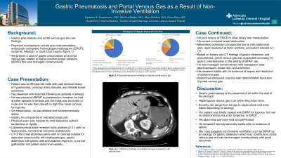
Case Description/Methods: An 86-year-old male with past medical history of hypertension, coronary artery disease, and irritable bowel syndrome presented with dyspnea after an episode of emesis. He was placed on BiPAP by paramedics; however, he had another episode of emesis and the mask was removed en route. He was then placed on high flow nasal cannula. On presentation, he was afebrile and hemodynamically stable. He reported mild abdominal pain initially. Mild distension without tenderness or rigidity was noted on exam. Laboratory evaluation revealed lactic acidosis of 3.1 with no leukocytosis, normal liver enzymes and bilirubin. Computed tomography (CT) chest abdomen pelvis with IV contrast revealed aspiration pneumonitis, left intrahepatic gas, gastric distension with gastric wall pneumatosis (Figure 1), a normal gallbladder and patent abdominal vessels. He had no history of ERCP or other biliary tree manipulation. He had no bowel obstruction. With mild abdominal pain, rapid resolution of lactic acidosis, and patent arteries on CT, mesenteric ischemia was not suspected. Based on history and CT findings of gastric distension and pneumatosis, portal venous gas was suspected due to gastric overdistension in the setting of barotrauma from BIPAP use. The patient was managed conservatively with nasogastric decompression, bowel rest, and antibiotics. He remained stable with no evidence of sepsis. His abdominal pain resolved. Abdominal ultrasound one day later demonstrated resolution of portal venous gas.
Discussion: Gastric pneumatosis is the presence of air within the wall of the stomach. Hepatoportal venous gas is air within the portal veins. Severity can range from benign to septic shock to even death depending on etiology. Our patient was briefly treated with BiPAP for hypoxia, but had no abdominal trauma, prior surgeries, or ERCP. His abdominal pain was mild and self-limited. He remained hemodynamically stable with no evidence of sepsis. Our case suggests non-invasive ventilation such as BiPAP as an etiology for gastric distension which may contribute to portal venous gas and can be managed conservatively with good outcomes.

Disclosures:
Madeline Sesselmann, DO1, Nikolina Madjer, MD2, Mary DeMent, DO3, Dean Silas, MD3. P1410 - Gastric Pneumatosis and Portal Venous Gas as a Result of Non-Invasive Ventilation, ACG 2023 Annual Scientific Meeting Abstracts. Vancouver, BC, Canada: American College of Gastroenterology.
1Advocate Aurora Health, Chicago, IL; 2Advocate Aurora Health, Park Ridge, IL; 3Advocate Aurora Health, Niles, IL
Introduction: Gastric pneumatosis and portal venous gas are rare findings on imaging. Proposed mechanisms include prior instrumentation, endoscopic retrograde cholangiopancreaticogram (ERCP), ischemia, infection, or blunt force trauma. We present a case of gastric pneumatosis and portal venous gas related to bilevel positive airway pressure (BiPAP) that was managed conservatively.
Case Description/Methods: An 86-year-old male with past medical history of hypertension, coronary artery disease, and irritable bowel syndrome presented with dyspnea after an episode of emesis. He was placed on BiPAP by paramedics; however, he had another episode of emesis and the mask was removed en route. He was then placed on high flow nasal cannula. On presentation, he was afebrile and hemodynamically stable. He reported mild abdominal pain initially. Mild distension without tenderness or rigidity was noted on exam. Laboratory evaluation revealed lactic acidosis of 3.1 with no leukocytosis, normal liver enzymes and bilirubin. Computed tomography (CT) chest abdomen pelvis with IV contrast revealed aspiration pneumonitis, left intrahepatic gas, gastric distension with gastric wall pneumatosis (Figure 1), a normal gallbladder and patent abdominal vessels. He had no history of ERCP or other biliary tree manipulation. He had no bowel obstruction. With mild abdominal pain, rapid resolution of lactic acidosis, and patent arteries on CT, mesenteric ischemia was not suspected. Based on history and CT findings of gastric distension and pneumatosis, portal venous gas was suspected due to gastric overdistension in the setting of barotrauma from BIPAP use. The patient was managed conservatively with nasogastric decompression, bowel rest, and antibiotics. He remained stable with no evidence of sepsis. His abdominal pain resolved. Abdominal ultrasound one day later demonstrated resolution of portal venous gas.
Discussion: Gastric pneumatosis is the presence of air within the wall of the stomach. Hepatoportal venous gas is air within the portal veins. Severity can range from benign to septic shock to even death depending on etiology. Our patient was briefly treated with BiPAP for hypoxia, but had no abdominal trauma, prior surgeries, or ERCP. His abdominal pain was mild and self-limited. He remained hemodynamically stable with no evidence of sepsis. Our case suggests non-invasive ventilation such as BiPAP as an etiology for gastric distension which may contribute to portal venous gas and can be managed conservatively with good outcomes.

Figure: Figure 1. CT abdomen pelvis with IV contrast demonstrating findings of portal venous gas, gastric dissension, and gastric wall pneumatosis.
Disclosures:
Madeline Sesselmann indicated no relevant financial relationships.
Nikolina Madjer indicated no relevant financial relationships.
Mary DeMent indicated no relevant financial relationships.
Dean Silas indicated no relevant financial relationships.
Madeline Sesselmann, DO1, Nikolina Madjer, MD2, Mary DeMent, DO3, Dean Silas, MD3. P1410 - Gastric Pneumatosis and Portal Venous Gas as a Result of Non-Invasive Ventilation, ACG 2023 Annual Scientific Meeting Abstracts. Vancouver, BC, Canada: American College of Gastroenterology.

