Sunday Poster Session
Category: Stomach
P1415 - T-Cell Lymphoma Mimicking as Lymphocytic Gastritis
Sunday, October 22, 2023
3:30 PM - 7:00 PM PT
Location: Exhibit Hall

Has Audio

Robert Freund, MD
University of Minnesota
Minneapolis, MN
Presenting Author(s)
Robert Freund, MD1, Kevin Turner, DO1, Holly Berg, DO1, Anna Pratt, MD1, Shawn Mallery, MD2, Michael Linden, MD, PhD1
1University of Minnesota, Minneapolis, MN; 2University of Minnesota Medical Center, Minneapolis, MN
Introduction: Lymphocytic gastritis (LG) is characterized by increased intraepithelial lymphocytes ( >25 per 100 epithelial cells). LG can be idiopathic or associated with Helicobacter, celiac disease, and various medications. Most gastric lymphomas are of B-cell origin and are composed of mucosal clusters of lymphocytes with enlarged nuclei. T-cell lymphomas can present with increased intraepithelial lymphocytes showing a histologic appearance nearly indistinguishable from LG; however, due to its rarity, T-cell lymphoma is rarely entertained as a differential. We present a CD8-positive T-cell lymphoma histologically resembling LG and highlight the importance of clinical history when reviewing gastric biopsies.
Case Description/Methods: A 62-year-old female with a history of monomorphic epitheliotropic intestinal T-cell lymphoma (MEITL) status post four cycles of CHOEP (cyclophosphamide, doxorubicin, etoposide, vincristine, and prednisone) chemotherapy presented with 4-5 days of post-prandial vomiting and solid-food dysphagia.
An esophagogastroduodenoscopy (EGD) was performed, and erythematous mucosa in the gastric body and antrum were biopsied. The reported endoscopic impression was likely reactive gastropathy.
The pathologist’s initial impression of the stomach biopsy was gastric mucosa with intraepithelial lymphocytosis, consistent with LG. Helicobacter was negative. Prior celiac serologies were negative for deamidated gliadin antibody IgG and IgA; and tissue transglutaminase antibodies IgG and IgA. However, a chart review revealed the patient’s history of a prior T-cell lymphoma of the small bowel.
A subsequent hematopathology consultation workup showed the atypical lymphoid infiltrate was composed of CD3 positive T-cells that were also positive for CD2, CD7 (bright), and CD8 while negative for CD4, CD5, and CD56. CD20 highlighted scattered B-cells.
A final diagnosis of recurrent/persistent CD8-positive mature T-cell lymphoma was made that was morphologically and immunohistochemically consistent with the patient’s prior MEITL.
Discussion: This case of CD8-positive mature T-cell lymphoma was recognized and diagnosed due to careful review of the patient’s chart. To our knowledge, only one other case of T-cell lymphoma presenting as morphologically-like lymphocytic gastritis has been described in the literature. Our case is also notable for the fact that the clinician impression during the EGD was likely reactive gastropathy, underscoring the importance of reviewing clinical history prior to final diagnosis.

Disclosures:
Robert Freund, MD1, Kevin Turner, DO1, Holly Berg, DO1, Anna Pratt, MD1, Shawn Mallery, MD2, Michael Linden, MD, PhD1. P1415 - T-Cell Lymphoma Mimicking as Lymphocytic Gastritis, ACG 2023 Annual Scientific Meeting Abstracts. Vancouver, BC, Canada: American College of Gastroenterology.
1University of Minnesota, Minneapolis, MN; 2University of Minnesota Medical Center, Minneapolis, MN
Introduction: Lymphocytic gastritis (LG) is characterized by increased intraepithelial lymphocytes ( >25 per 100 epithelial cells). LG can be idiopathic or associated with Helicobacter, celiac disease, and various medications. Most gastric lymphomas are of B-cell origin and are composed of mucosal clusters of lymphocytes with enlarged nuclei. T-cell lymphomas can present with increased intraepithelial lymphocytes showing a histologic appearance nearly indistinguishable from LG; however, due to its rarity, T-cell lymphoma is rarely entertained as a differential. We present a CD8-positive T-cell lymphoma histologically resembling LG and highlight the importance of clinical history when reviewing gastric biopsies.
Case Description/Methods: A 62-year-old female with a history of monomorphic epitheliotropic intestinal T-cell lymphoma (MEITL) status post four cycles of CHOEP (cyclophosphamide, doxorubicin, etoposide, vincristine, and prednisone) chemotherapy presented with 4-5 days of post-prandial vomiting and solid-food dysphagia.
An esophagogastroduodenoscopy (EGD) was performed, and erythematous mucosa in the gastric body and antrum were biopsied. The reported endoscopic impression was likely reactive gastropathy.
The pathologist’s initial impression of the stomach biopsy was gastric mucosa with intraepithelial lymphocytosis, consistent with LG. Helicobacter was negative. Prior celiac serologies were negative for deamidated gliadin antibody IgG and IgA; and tissue transglutaminase antibodies IgG and IgA. However, a chart review revealed the patient’s history of a prior T-cell lymphoma of the small bowel.
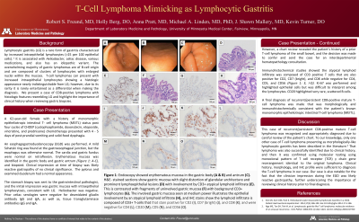
A subsequent hematopathology consultation workup showed the atypical lymphoid infiltrate was composed of CD3 positive T-cells that were also positive for CD2, CD7 (bright), and CD8 while negative for CD4, CD5, and CD56. CD20 highlighted scattered B-cells.
A final diagnosis of recurrent/persistent CD8-positive mature T-cell lymphoma was made that was morphologically and immunohistochemically consistent with the patient’s prior MEITL.
Discussion: This case of CD8-positive mature T-cell lymphoma was recognized and diagnosed due to careful review of the patient’s chart. To our knowledge, only one other case of T-cell lymphoma presenting as morphologically-like lymphocytic gastritis has been described in the literature. Our case is also notable for the fact that the clinician impression during the EGD was likely reactive gastropathy, underscoring the importance of reviewing clinical history prior to final diagnosis.

Figure: Figure 1: Endoscopy showed erythematous mucosa in the gastric body (A & B) and antrum (C). H&E-stained sections show gastric mucosa with slight distortion of glandular architecture and prominent lymphoepithelial lesions (D) with involvement by a CD3+ atypical lymphoid infiltrate (E). This is contrasted with fragments of uninvolved gastric mucosa (F) with background CD3+ lymphocytes (G). The involved gastric mucosa seen at medium power illustrates the epithelial involvement by an atypical lymphoid infiltrate (H), and IHC stains show the lymphoid infiltrate is composed of CD3+ T-cells that stain positive for CD2 (I), CD7 (bright) (J), and CD8 (K); and were negative for CD4 (L), CD20 (M), CD5 (N), and CD56 (O).
Disclosures:
Robert Freund indicated no relevant financial relationships.
Kevin Turner indicated no relevant financial relationships.
Holly Berg indicated no relevant financial relationships.
Anna Pratt indicated no relevant financial relationships.
Shawn Mallery indicated no relevant financial relationships.
Michael Linden indicated no relevant financial relationships.
Robert Freund, MD1, Kevin Turner, DO1, Holly Berg, DO1, Anna Pratt, MD1, Shawn Mallery, MD2, Michael Linden, MD, PhD1. P1415 - T-Cell Lymphoma Mimicking as Lymphocytic Gastritis, ACG 2023 Annual Scientific Meeting Abstracts. Vancouver, BC, Canada: American College of Gastroenterology.

