Sunday Poster Session
Category: Stomach
P1382 - I Spy With My Owl's Eye: Gastrointestinal Cytomegalovirus
Sunday, October 22, 2023
3:30 PM - 7:00 PM PT
Location: Exhibit Hall

Has Audio

Rita Rehana, MD
Ascension Macomb-Oakland
Peoria, AZ
Presenting Author(s)
Rita Rehana, MD1, Achyut Bhattacharyya, MD2, Alex Heimbigner, MD2, Stephen Hoffman, DO3
1Ascension Macomb-Oakland, Peoria, AZ; 2Banner University Medical Center, Tucson, AZ; 3Ascension Macomb-Oakland, Warren, MI
Introduction: Cytomegalovirus (CMV) is an opportunistic pathogen among immunocompromised hosts. Individuals with human immunodeficiency virus, immunosuppressive states, and long-term steroid use are most susceptible. CMV infection can affect several organs, but gastrointestinal (GI) involvement is one of the most common. GI CMV manifestations include nonspecific symptomatology such as dysphagia, abdominal pain, diarrhea, and GI bleeding. However, CMV gastritis is rare with only a few hundred cases described in literature. We describe a case of an immunocompromised man presenting with symptomatic anemia.
Case Description/Methods: A 78-year-old gentleman with diffuse large B-cell lymphoma and peritoneal carcinomatosis on chemotherapy presented with complaints of dizziness. Initially, vitals showed blood pressure (BP) 84/44 and heart rate 125. He was found to have pancytopenia with hemoglobin 5.9 and other electrolyte derangements. He received a blood transfusion and electrolyte replacement with improvement in BP and symptoms. He denied overt bleeding. However, during his hospitalization, he experienced multiple episodes of hematochezia requiring another blood transfusion. CT angiogram did not reveal any evidence of active extravasation or intraabdominal/pelvic abnormalities. Gastroenterology was consulted for a bidirectional endoscopy which revealed many nonbleeding ulcers with a clean ulcer base in the gastric body. Biopsies of the numerous areas of patchy erythema in the gastric body were obtained. Histopathological examination showed large epithelial cells with a characteristic “owl’s eye” eosinophilic intranuclear inclusion body surrounded by a clear halo. Immunostaining for CMV antigens were positive. The endoscopic and histopathological findings were consistent with CMV gastritis. Gastric lymphoma and Helicobacter pylori were negative.
Discussion: Data specific to GI CMV infections in immunocompromised patients is still limited, likely due to disease under recognition, which could lead to diagnostic delay and poor outcomes. As depicted in our patient, the “owl’s eye” intranuclear inclusions are the hallmark of CMV infection. Ultimately, endoscopic findings are variable and nondiagnostic macroscopically. Therefore, determination of a diagnosis is dependent on pathology and serology. Hence, several diagnostic tools must be utilized, such as CMV antigenemia assay and polymerase chain reaction. Overall, most GI CMV infections respond well to ganciclovir despite the underlying cause of immunosuppression.

Disclosures:
Rita Rehana, MD1, Achyut Bhattacharyya, MD2, Alex Heimbigner, MD2, Stephen Hoffman, DO3. P1382 - I Spy With My Owl's Eye: Gastrointestinal Cytomegalovirus, ACG 2023 Annual Scientific Meeting Abstracts. Vancouver, BC, Canada: American College of Gastroenterology.
1Ascension Macomb-Oakland, Peoria, AZ; 2Banner University Medical Center, Tucson, AZ; 3Ascension Macomb-Oakland, Warren, MI
Introduction: Cytomegalovirus (CMV) is an opportunistic pathogen among immunocompromised hosts. Individuals with human immunodeficiency virus, immunosuppressive states, and long-term steroid use are most susceptible. CMV infection can affect several organs, but gastrointestinal (GI) involvement is one of the most common. GI CMV manifestations include nonspecific symptomatology such as dysphagia, abdominal pain, diarrhea, and GI bleeding. However, CMV gastritis is rare with only a few hundred cases described in literature. We describe a case of an immunocompromised man presenting with symptomatic anemia.
Case Description/Methods: A 78-year-old gentleman with diffuse large B-cell lymphoma and peritoneal carcinomatosis on chemotherapy presented with complaints of dizziness. Initially, vitals showed blood pressure (BP) 84/44 and heart rate 125. He was found to have pancytopenia with hemoglobin 5.9 and other electrolyte derangements. He received a blood transfusion and electrolyte replacement with improvement in BP and symptoms. He denied overt bleeding. However, during his hospitalization, he experienced multiple episodes of hematochezia requiring another blood transfusion. CT angiogram did not reveal any evidence of active extravasation or intraabdominal/pelvic abnormalities. Gastroenterology was consulted for a bidirectional endoscopy which revealed many nonbleeding ulcers with a clean ulcer base in the gastric body. Biopsies of the numerous areas of patchy erythema in the gastric body were obtained. Histopathological examination showed large epithelial cells with a characteristic “owl’s eye” eosinophilic intranuclear inclusion body surrounded by a clear halo. Immunostaining for CMV antigens were positive. The endoscopic and histopathological findings were consistent with CMV gastritis. Gastric lymphoma and Helicobacter pylori were negative.
Discussion: Data specific to GI CMV infections in immunocompromised patients is still limited, likely due to disease under recognition, which could lead to diagnostic delay and poor outcomes. As depicted in our patient, the “owl’s eye” intranuclear inclusions are the hallmark of CMV infection. Ultimately, endoscopic findings are variable and nondiagnostic macroscopically. Therefore, determination of a diagnosis is dependent on pathology and serology. Hence, several diagnostic tools must be utilized, such as CMV antigenemia assay and polymerase chain reaction. Overall, most GI CMV infections respond well to ganciclovir despite the underlying cause of immunosuppression.

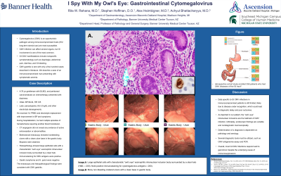
Figure: Histopathological and endoscopic pictures of cytomegalovirus gastritis. Note many non-bleeding cratered gastric ulcers with a clean ulcer base (Forrest Class III) and numerous areas of patchy erythema in the gastric body; Histopathological examination of the erythema showing large epithelial cells with characteristic “owl’s eye” (Arrow) eosinophilic intranuclear inclusion body surrounded by a clear halo (H&E, × 200); (positive immunostaining for cytomegalovirus antigens × 200).
Disclosures:
Rita Rehana indicated no relevant financial relationships.
Achyut Bhattacharyya indicated no relevant financial relationships.
Alex Heimbigner indicated no relevant financial relationships.
Stephen Hoffman indicated no relevant financial relationships.
Rita Rehana, MD1, Achyut Bhattacharyya, MD2, Alex Heimbigner, MD2, Stephen Hoffman, DO3. P1382 - I Spy With My Owl's Eye: Gastrointestinal Cytomegalovirus, ACG 2023 Annual Scientific Meeting Abstracts. Vancouver, BC, Canada: American College of Gastroenterology.
