Sunday Poster Session
Category: Liver
P1155 - A Case of Inflammatory Pseudotumor of the Liver Associated With Influenza A
Sunday, October 22, 2023
3:30 PM - 7:00 PM PT
Location: Exhibit Hall

Has Audio
- AC
Alexander Chen, MD
Rutgers Robert Wood Johnson Medical School
New Brunswick, New Jersey
Presenting Author(s)
Ankoor H. Patel, MD, Alexander Chen, MD, Alexander Lalos, MD
Rutgers Robert Wood Johnson Medical School, New Brunswick, NJ
Introduction: Inflammatory pseudotumor (IPT) is a rare, benign tumor which can mimic malignancy. We report a case of hepatic IPT following diagnosis of influenza A, an association that has not been previously reported.
Case Description/Methods: A 65-year-old male with history of type 2 diabetes and obesity was referred to hepatology clinic for evaluation of multiple liver masses. Five months prior he reported fevers, confusion and unintentional weight loss of 40lbs in a 3-week timespan. He was seen in the emergency department of an outside hospital and a workup was positive for influenza A, diagnosed via nasal swab, as well as a CRP of 191.0mg/L. He was treated with oseltamivir. Because the patient's symptoms were persisting his primary care physician obtained a CT scan which revealed three heterogeneous left hepatic lobe masses and one hypodense right hepatic lobe mass with adenopathy of the periportal, paraceliac and mediastinal regions. A subsequent positron emission tomography (PET) scan showed small focal areas of uptake but overall uptake of the left lobe hepatic masses appeared similar to the adjacent normal liver. At this time, the patient's symptoms began to improve. Fine need aspiration (FNA) of the masses revealed mixed inflammatory infiltrate but no malignant cells. A core biopsy was then obtained. Trichrome stain highlighted dense fibrosis. CK7 immunostain showed preserved bile ducts and smooth muscle actin immunostain showed extensive reactivity in the fibrous stroma. Immunostains for IgG4 and ALK1 were negative. Findings were felt to be consistent with hepatic IPT by the pathologist. When seen in our clinic the patient reported resolution of his symptoms. Repeat CT revealed a significant decrease in size of the left hepatic lobe masses and resolution of the right hepatic lobe mass. The degree of adenopathy was decreased or stable. An upper endoscopy and colonoscopy revealed only gastritis and two small benign rectal polyps. Repeat lab work showed a decrease in CRP to 2mg/L. Ca19-9 was 42 mg/dL, AFP and CEA were normal. Hepatitis C and B serologies were all non-reactive as were autoimmune hepatitis serologies. The final diagnosis was IPT and the patient has since returned to his baseline health.
Discussion: We present the first case of hepatic IPT in a patient with recently diagnosed influenza A. Our case highlights the importance of including IPT in the differential diagnosis in a patient who presents with clinical and radiologic findings suspicious for hepatic malignancy following influenza A.

Disclosures:
Ankoor H. Patel, MD, Alexander Chen, MD, Alexander Lalos, MD. P1155 - A Case of Inflammatory Pseudotumor of the Liver Associated With Influenza A, ACG 2023 Annual Scientific Meeting Abstracts. Vancouver, BC, Canada: American College of Gastroenterology.
Rutgers Robert Wood Johnson Medical School, New Brunswick, NJ
Introduction: Inflammatory pseudotumor (IPT) is a rare, benign tumor which can mimic malignancy. We report a case of hepatic IPT following diagnosis of influenza A, an association that has not been previously reported.
Case Description/Methods: A 65-year-old male with history of type 2 diabetes and obesity was referred to hepatology clinic for evaluation of multiple liver masses. Five months prior he reported fevers, confusion and unintentional weight loss of 40lbs in a 3-week timespan. He was seen in the emergency department of an outside hospital and a workup was positive for influenza A, diagnosed via nasal swab, as well as a CRP of 191.0mg/L. He was treated with oseltamivir. Because the patient's symptoms were persisting his primary care physician obtained a CT scan which revealed three heterogeneous left hepatic lobe masses and one hypodense right hepatic lobe mass with adenopathy of the periportal, paraceliac and mediastinal regions. A subsequent positron emission tomography (PET) scan showed small focal areas of uptake but overall uptake of the left lobe hepatic masses appeared similar to the adjacent normal liver. At this time, the patient's symptoms began to improve. Fine need aspiration (FNA) of the masses revealed mixed inflammatory infiltrate but no malignant cells. A core biopsy was then obtained. Trichrome stain highlighted dense fibrosis. CK7 immunostain showed preserved bile ducts and smooth muscle actin immunostain showed extensive reactivity in the fibrous stroma. Immunostains for IgG4 and ALK1 were negative. Findings were felt to be consistent with hepatic IPT by the pathologist. When seen in our clinic the patient reported resolution of his symptoms. Repeat CT revealed a significant decrease in size of the left hepatic lobe masses and resolution of the right hepatic lobe mass. The degree of adenopathy was decreased or stable. An upper endoscopy and colonoscopy revealed only gastritis and two small benign rectal polyps. Repeat lab work showed a decrease in CRP to 2mg/L. Ca19-9 was 42 mg/dL, AFP and CEA were normal. Hepatitis C and B serologies were all non-reactive as were autoimmune hepatitis serologies. The final diagnosis was IPT and the patient has since returned to his baseline health.
Discussion: We present the first case of hepatic IPT in a patient with recently diagnosed influenza A. Our case highlights the importance of including IPT in the differential diagnosis in a patient who presents with clinical and radiologic findings suspicious for hepatic malignancy following influenza A.

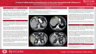
Figure: (A) A 5.0 cm x 3.0 cm mildly hypodense hepatic mass on CT A/P dual phase liver. (B) The same mass 3 months later, now 1.9 cm x 1.3 cm on CT A/P dual phase liver.
Disclosures:
Ankoor Patel indicated no relevant financial relationships.
Alexander Chen indicated no relevant financial relationships.
Alexander Lalos indicated no relevant financial relationships.
Ankoor H. Patel, MD, Alexander Chen, MD, Alexander Lalos, MD. P1155 - A Case of Inflammatory Pseudotumor of the Liver Associated With Influenza A, ACG 2023 Annual Scientific Meeting Abstracts. Vancouver, BC, Canada: American College of Gastroenterology.
