Sunday Poster Session
Category: Esophagus
P0492 - Luck of the Draw: Incidental Biopsy With High-Grade Dysplasia in the Setting of Lymphocytic Esophagitis
Sunday, October 22, 2023
3:30 PM - 7:00 PM PT
Location: Exhibit Hall

Has Audio

Bharat Ponnaganti, MD
University of South Florida
Tampa, Florida
Presenting Author(s)
Bharat Ponnaganti, MD1, Krishen Patel, MD2, John Jacobs, MD3
1University of South Florida, Tampa, FL; 2Unviersity of South Florida, Tampa, FL; 3University of South Florida Morsani College of Medicine, Tampa, FL
Introduction: Lymphocytic esophagitis (LyE) is a rare form of chronic esophagitis with poorly understood pathogenesis and prognosis. Literature is scarce on the potential associations between LyE and dysplasia as well as the long-term clinical outcomes of patients with LyE. We present a case of an elderly female with known LyE found to have a 2cm segment of high-grade squamous dysplasia on esophageal biopsy.
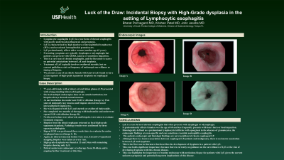
Case Description/Methods: Patient is a 77-year-old female with a history of oral lichen planus (LP) presented with a long-standing history of dysphagia. She had multiple outside endoscopies in the past with normal biopsies.She then underwent EGD with dilation (Image A). EGD showed minimally lacy mucosa and esophageal biopsies showed LyE. She was treated with swallowed Budesonide. After two months of therapy, she underwent repeat EGD for further esophageal dilation. At that time, while no discreet mucosal lesions were seen in the entire esophagus (Image B), repeat biopsies were taken to reevaluate for response to therapy. Biopsies from the distal esophagus showed focal high-grade squamous dysplasia. Repeat EGD was performed three weeks later to re-evaluate the entire esophageal mucosa (Image C,D). Again, no discreet mucosal lesions were seen. Extensive 4-quadrant mapping biopsies were taken every 1 cm. High-grade dysplasia was found at 33 and 34cm with remaining biopsies showing only LyE. Patient underwent endoscopic cryotherapy from 30-40cm and is ongoing further treatment at this time.
Discussion: LyE is a rare disease with patients often presenting with dysphagia. Predominantly affects females above the age of 60 and less frequently causes heartburn or food impaction. Histologically defined as a predominant lymphocytic infiltrate with spongiosis in the absence of granulocytes, the endoscopic findings are non-specific and can sometimes resemble eosinophilic esophagitis. This patients endoscopic and histologic findings are not classic for esophageal LP. While patients with esophageal LP have a higher risk of malignancy, this is the first case in literature that describes the development of dysplasia in a patient with LyE. Furthermore, guidance on the surveillance of LyE and the risk of developing dysplasia does not exist. The current mainstay of treatment for LyE includes acid suppressive therapy and swallowed steroids. This case highlights the importance of routine endoscopy with meticulous biopsy for patients with LyE given the current unknown prognosis and long-term implications of this rare disease.

Disclosures:
Bharat Ponnaganti, MD1, Krishen Patel, MD2, John Jacobs, MD3. P0492 - Luck of the Draw: Incidental Biopsy With High-Grade Dysplasia in the Setting of Lymphocytic Esophagitis, ACG 2023 Annual Scientific Meeting Abstracts. Vancouver, BC, Canada: American College of Gastroenterology.
1University of South Florida, Tampa, FL; 2Unviersity of South Florida, Tampa, FL; 3University of South Florida Morsani College of Medicine, Tampa, FL
Introduction: Lymphocytic esophagitis (LyE) is a rare form of chronic esophagitis with poorly understood pathogenesis and prognosis. Literature is scarce on the potential associations between LyE and dysplasia as well as the long-term clinical outcomes of patients with LyE. We present a case of an elderly female with known LyE found to have a 2cm segment of high-grade squamous dysplasia on esophageal biopsy.
Case Description/Methods: Patient is a 77-year-old female with a history of oral lichen planus (LP) presented with a long-standing history of dysphagia. She had multiple outside endoscopies in the past with normal biopsies.She then underwent EGD with dilation (Image A). EGD showed minimally lacy mucosa and esophageal biopsies showed LyE. She was treated with swallowed Budesonide. After two months of therapy, she underwent repeat EGD for further esophageal dilation. At that time, while no discreet mucosal lesions were seen in the entire esophagus (Image B), repeat biopsies were taken to reevaluate for response to therapy. Biopsies from the distal esophagus showed focal high-grade squamous dysplasia. Repeat EGD was performed three weeks later to re-evaluate the entire esophageal mucosa (Image C,D). Again, no discreet mucosal lesions were seen. Extensive 4-quadrant mapping biopsies were taken every 1 cm. High-grade dysplasia was found at 33 and 34cm with remaining biopsies showing only LyE. Patient underwent endoscopic cryotherapy from 30-40cm and is ongoing further treatment at this time.
Discussion: LyE is a rare disease with patients often presenting with dysphagia. Predominantly affects females above the age of 60 and less frequently causes heartburn or food impaction. Histologically defined as a predominant lymphocytic infiltrate with spongiosis in the absence of granulocytes, the endoscopic findings are non-specific and can sometimes resemble eosinophilic esophagitis. This patients endoscopic and histologic findings are not classic for esophageal LP. While patients with esophageal LP have a higher risk of malignancy, this is the first case in literature that describes the development of dysplasia in a patient with LyE. Furthermore, guidance on the surveillance of LyE and the risk of developing dysplasia does not exist. The current mainstay of treatment for LyE includes acid suppressive therapy and swallowed steroids. This case highlights the importance of routine endoscopy with meticulous biopsy for patients with LyE given the current unknown prognosis and long-term implications of this rare disease.

Figure: Images of the distal esophagus from multiple Esophagogastroduodenoscopies
Disclosures:
Bharat Ponnaganti indicated no relevant financial relationships.
Krishen Patel indicated no relevant financial relationships.
John Jacobs indicated no relevant financial relationships.
Bharat Ponnaganti, MD1, Krishen Patel, MD2, John Jacobs, MD3. P0492 - Luck of the Draw: Incidental Biopsy With High-Grade Dysplasia in the Setting of Lymphocytic Esophagitis, ACG 2023 Annual Scientific Meeting Abstracts. Vancouver, BC, Canada: American College of Gastroenterology.
