Sunday Poster Session
Category: Esophagus
P0501 - A Large Esophageal Mass With Intramural Esophageal Dissection: An Unusual Presentation of Herpes Simplex Esophagitis
Sunday, October 22, 2023
3:30 PM - 7:00 PM PT
Location: Exhibit Hall

Has Audio

Azizullah Beran, MD
Indiana University
Indianapolis, IN
Presenting Author(s)
Azizullah Beran, MD1, Ramzi Hassouneh, DO1, Eleazar E.. Montalvan-Sanchez, MD2, Mohammad Al-Haddad, MD1, Zhibo An, MD1
1Indiana University, Indianapolis, IN; 2Indiana University School of Medicine, Indianapolis, IN
Introduction: Herpes simplex virus (HSV) esophagitis primarily affects immunocompromised individuals, displaying characteristic scattered "volcano-appearing" ulcers typically smaller than 2 cm on endoscopy. Uncommon presentations include exudates, plaques, or diffuse erosive esophagitis. To date, no cases of HSV esophagitis presenting as an esophageal mass have been reported. Here, we present a rare case of HSV esophagitis mimicking an esophageal mass in a patient with metastatic lung cancer receiving a high-dose steroid therapy.
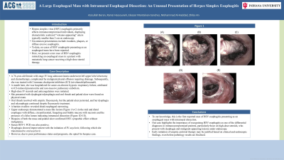
Case Description/Methods: A 76-year-old female with stage IV lung adenocarcinoma underwent left upper lobe lobectomy and chemotherapy, complicated by malignant pleural effusion requiring drainage. Subsequently, she was treated with 2 immune checkpoint inhibitors (ICI) (nivolumab/ipilimumab). A month later, she was hospitalized for acute-on-chronic hypoxic respiratory failure, attributed to ICI-induced pneumonitis and non-massive pulmonary embolism. High-dose IV steroids and anticoagulation were initiated. She presented with dysphagia/odynophagia and oral thrush and palatal ulcer were found on physical exam. Oral thrush resolved with empiric fluconazole, but the palatal ulcer persisted, and her dysphagia and odynophagia continued despite fluconazole treatment. A barium swallow revealed distal esophageal narrowing. Upper endoscopy demonstrated a mass-like lesion (Figure 1A-C) in the mid and distal esophagus with diffuse, circumferential, fungating and friable mucosa with necrosis and the presence of a false lumen indicating intramural dissection (Figure 1D-1E). Biopsies of both the mass and palatal ulcer confirmed HSV cytopathic effect without malignancy. Serum HSV-1 PCR was also positive. Dysphagia showed improvement with the initiation of IV acyclovir, following which she transitioned to oral acyclovir. However, due to poor performance status and prognosis, she opted for hospice care.
Discussion: To our knowledge, this is the first reported case of HSV esophagitis presenting as an esophageal mass with intramural dissection. Our case highlights the importance of recognizing HSV esophagitis as one of the differential diagnoses in immunocompromised patients, particularly those on high-dose steroids, who present with dysphagia and malignant appearing lesions under endoscopy. Early initiation of empiric antiviral therapy may be justified based on clinical and endoscopic findings, even before pathology results are finalized.

Disclosures:
Azizullah Beran, MD1, Ramzi Hassouneh, DO1, Eleazar E.. Montalvan-Sanchez, MD2, Mohammad Al-Haddad, MD1, Zhibo An, MD1. P0501 - A Large Esophageal Mass With Intramural Esophageal Dissection: An Unusual Presentation of Herpes Simplex Esophagitis, ACG 2023 Annual Scientific Meeting Abstracts. Vancouver, BC, Canada: American College of Gastroenterology.
1Indiana University, Indianapolis, IN; 2Indiana University School of Medicine, Indianapolis, IN
Introduction: Herpes simplex virus (HSV) esophagitis primarily affects immunocompromised individuals, displaying characteristic scattered "volcano-appearing" ulcers typically smaller than 2 cm on endoscopy. Uncommon presentations include exudates, plaques, or diffuse erosive esophagitis. To date, no cases of HSV esophagitis presenting as an esophageal mass have been reported. Here, we present a rare case of HSV esophagitis mimicking an esophageal mass in a patient with metastatic lung cancer receiving a high-dose steroid therapy.
Case Description/Methods: A 76-year-old female with stage IV lung adenocarcinoma underwent left upper lobe lobectomy and chemotherapy, complicated by malignant pleural effusion requiring drainage. Subsequently, she was treated with 2 immune checkpoint inhibitors (ICI) (nivolumab/ipilimumab). A month later, she was hospitalized for acute-on-chronic hypoxic respiratory failure, attributed to ICI-induced pneumonitis and non-massive pulmonary embolism. High-dose IV steroids and anticoagulation were initiated. She presented with dysphagia/odynophagia and oral thrush and palatal ulcer were found on physical exam. Oral thrush resolved with empiric fluconazole, but the palatal ulcer persisted, and her dysphagia and odynophagia continued despite fluconazole treatment. A barium swallow revealed distal esophageal narrowing. Upper endoscopy demonstrated a mass-like lesion (Figure 1A-C) in the mid and distal esophagus with diffuse, circumferential, fungating and friable mucosa with necrosis and the presence of a false lumen indicating intramural dissection (Figure 1D-1E). Biopsies of both the mass and palatal ulcer confirmed HSV cytopathic effect without malignancy. Serum HSV-1 PCR was also positive. Dysphagia showed improvement with the initiation of IV acyclovir, following which she transitioned to oral acyclovir. However, due to poor performance status and prognosis, she opted for hospice care.
Discussion: To our knowledge, this is the first reported case of HSV esophagitis presenting as an esophageal mass with intramural dissection. Our case highlights the importance of recognizing HSV esophagitis as one of the differential diagnoses in immunocompromised patients, particularly those on high-dose steroids, who present with dysphagia and malignant appearing lesions under endoscopy. Early initiation of empiric antiviral therapy may be justified based on clinical and endoscopic findings, even before pathology results are finalized.

Figure: Figure 1
Disclosures:
Azizullah Beran indicated no relevant financial relationships.
Ramzi Hassouneh indicated no relevant financial relationships.
Eleazar Montalvan-Sanchez indicated no relevant financial relationships.
Mohammad Al-Haddad: Amplified Sciences – Grant/Research Support. Creatics, LLC – Grant/Research Support. Interpace diagnostics – Consultant.
Zhibo An indicated no relevant financial relationships.
Azizullah Beran, MD1, Ramzi Hassouneh, DO1, Eleazar E.. Montalvan-Sanchez, MD2, Mohammad Al-Haddad, MD1, Zhibo An, MD1. P0501 - A Large Esophageal Mass With Intramural Esophageal Dissection: An Unusual Presentation of Herpes Simplex Esophagitis, ACG 2023 Annual Scientific Meeting Abstracts. Vancouver, BC, Canada: American College of Gastroenterology.
