Monday Poster Session
Category: Esophagus
P1876 - Dysphagia Lusoria in the Setting of CLOVES Syndrome
Monday, October 23, 2023
10:30 AM - 4:15 PM PT
Location: Exhibit Hall

Has Audio
- CB
Christopher Bouvette, MD
University of Oklahoma College of Medicine
Oklahoma City, Oklahoma
Presenting Author(s)
Christopher Bouvette, MD, Ahmad Basil Nasir, MD, Michael Sutton, MD, Ijlal A. Ali, MD
University of Oklahoma College of Medicine, Oklahoma City, OK
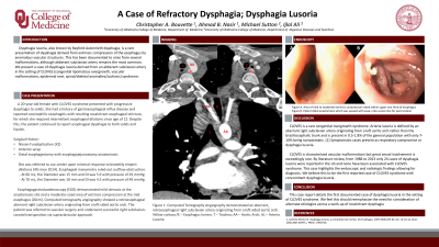
Introduction: Dysphagia lusoria, also known by Bayford-Autenrieth dysphagia, is a rare presentation of dysphagia derived from extrinsic compression of the esophagus by anomalous vascular structures. We present a case of dysphagia lusoria derived from an abberant subclavian artery in the setting of CLOVES (congenital lipomatous overgrowth, vascular malformations, epidermal nevi, spinal/skeletal anomalies/scoliosis) syndrome.
Case Description/Methods: A 20-year-old female with CLOVES syndrome presented with progressive dysphagia to solids. She had a history of gastroesophageal reflux disease and reported eosinophilic esophagitis with resulting recalcitrant esophageal stricture. She required intermittent esophageal dilations since age of 12 and underwent Nissen Fundoplication, followed by anterior wrap, and upon failure distal esophagectomy with esophagojejunostomy anastomosis. Despite this, the patient continued to report esophageal dysphagia to both solids and liquids.
She was referred to our center upon minimal response to biweekly empiric dilations. Esophageal manometry ruled-out outflow obstruction. Esophagogastroduodenoscopy (EGD) demonstrated mild stenosis at the anastomosis site and a moderate-sized area of extrinsic compression at the mid esophagus (20cm). Computed tomography angiography showed a retroesophageal aberrant right subclavian artery originating from a left-sided aortic arch. The patient was referred to vascular surgery and underwent successful right subclavian-carotid transposition via supraclavicular approach.
Discussion: CLOVES is a rare congenital overgrowth syndrome. Arteria lusoria is defined by an aberrant right subclavian artery originating from a left aortic arch rather than the brachiocephalic trunk and is present in 0.5-1.8% of the general population with only 7-10% being symptomatic. [1] Symptomatic cases present as respiratory compromise or dysphagia lusoria.
CLOVES is characterized vascular malformations but great vessel involvement is exceedingly rare. By literature review, from 1988 to 2013 only 24 cases of dysphagia lusoria were reported in the US and none have been associated with CLOVES syndrome. This case highlights the endoscopic and radiologic findings allowing for diagnosis. We believe this to be the first reported case of CLOVES syndrome with concomitant dysphagia lusoria.
1. Levitt B, Richter JE. Dysphagia lusoria: a comprehensive review. Dis Esophagus. 2007;20(6):455-60. doi: 10.1111/j.1442-2050.2007.00787.x. PMID: 17958718.

Disclosures:
Christopher Bouvette, MD, Ahmad Basil Nasir, MD, Michael Sutton, MD, Ijlal A. Ali, MD. P1876 - Dysphagia Lusoria in the Setting of CLOVES Syndrome, ACG 2023 Annual Scientific Meeting Abstracts. Vancouver, BC, Canada: American College of Gastroenterology.
University of Oklahoma College of Medicine, Oklahoma City, OK
Introduction: Dysphagia lusoria, also known by Bayford-Autenrieth dysphagia, is a rare presentation of dysphagia derived from extrinsic compression of the esophagus by anomalous vascular structures. We present a case of dysphagia lusoria derived from an abberant subclavian artery in the setting of CLOVES (congenital lipomatous overgrowth, vascular malformations, epidermal nevi, spinal/skeletal anomalies/scoliosis) syndrome.
Case Description/Methods: A 20-year-old female with CLOVES syndrome presented with progressive dysphagia to solids. She had a history of gastroesophageal reflux disease and reported eosinophilic esophagitis with resulting recalcitrant esophageal stricture. She required intermittent esophageal dilations since age of 12 and underwent Nissen Fundoplication, followed by anterior wrap, and upon failure distal esophagectomy with esophagojejunostomy anastomosis. Despite this, the patient continued to report esophageal dysphagia to both solids and liquids.
She was referred to our center upon minimal response to biweekly empiric dilations. Esophageal manometry ruled-out outflow obstruction. Esophagogastroduodenoscopy (EGD) demonstrated mild stenosis at the anastomosis site and a moderate-sized area of extrinsic compression at the mid esophagus (20cm). Computed tomography angiography showed a retroesophageal aberrant right subclavian artery originating from a left-sided aortic arch. The patient was referred to vascular surgery and underwent successful right subclavian-carotid transposition via supraclavicular approach.
Discussion: CLOVES is a rare congenital overgrowth syndrome. Arteria lusoria is defined by an aberrant right subclavian artery originating from a left aortic arch rather than the brachiocephalic trunk and is present in 0.5-1.8% of the general population with only 7-10% being symptomatic. [1] Symptomatic cases present as respiratory compromise or dysphagia lusoria.
CLOVES is characterized vascular malformations but great vessel involvement is exceedingly rare. By literature review, from 1988 to 2013 only 24 cases of dysphagia lusoria were reported in the US and none have been associated with CLOVES syndrome. This case highlights the endoscopic and radiologic findings allowing for diagnosis. We believe this to be the first reported case of CLOVES syndrome with concomitant dysphagia lusoria.
1. Levitt B, Richter JE. Dysphagia lusoria: a comprehensive review. Dis Esophagus. 2007;20(6):455-60. doi: 10.1111/j.1442-2050.2007.00787.x. PMID: 17958718.

Figure: Figure 1.
Computed Tomography Angiography demonstrated an aberrant, retroesophageal right subclavian artery originating from a left-sided aortic arch.
Yellow contour/E – Esophagus lumen; T – Trachea; AA – Aortic Arch; AL – Arteria Lusoria
Computed Tomography Angiography demonstrated an aberrant, retroesophageal right subclavian artery originating from a left-sided aortic arch.
Yellow contour/E – Esophagus lumen; T – Trachea; AA – Aortic Arch; AL – Arteria Lusoria
Disclosures:
Christopher Bouvette indicated no relevant financial relationships.
Ahmad Basil Nasir indicated no relevant financial relationships.
Michael Sutton indicated no relevant financial relationships.
Ijlal Ali indicated no relevant financial relationships.
Christopher Bouvette, MD, Ahmad Basil Nasir, MD, Michael Sutton, MD, Ijlal A. Ali, MD. P1876 - Dysphagia Lusoria in the Setting of CLOVES Syndrome, ACG 2023 Annual Scientific Meeting Abstracts. Vancouver, BC, Canada: American College of Gastroenterology.
