Monday Poster Session
Category: Esophagus
P1890 - Traction Diverticula in Patients with Pulmonary Granulomatous Disease
Monday, October 23, 2023
10:30 AM - 4:15 PM PT
Location: Exhibit Hall

Has Audio
- CX
Chelsea Xu, MD
University of California, San Francisco
San Francisco, California
Presenting Author(s)
Chelsea Xu, MD, Myung Ko, MD, Priya Kathpalia, MD
University of California, San Francisco, San Francisco, CA
Introduction: Mid-esophageal diverticula account for < 30% of esophageal diverticula. They often result from traction caused by chronic mediastinal inflammation, leading to full-thickness esophageal wall herniation. We present four cases of traction diverticula at a single institution.
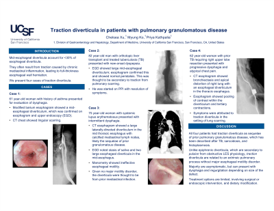
Case Description/Methods: In Case 1, a 61 year-old woman with history of asthma presented for evaluation of dysphagia. A modified barium esophagram noted a mid-esophageal diverticulum, which was confirmed on esophagram and upper endoscopy (EGD) (A, B). CT chest showed lingular scarring. In Case 2, an 82 year-old man with orthotopic liver transplant and treated tuberculosis (TB) presented with new-onset dyspepsia. EGD showed large mid-esophageal diverticula; esophagram confirmed this and showed normal peristalsis (C). This was thought to be secondary to traction from pulmonary scarring. He was started on PPI with resolution of symptoms. In Case 3, a 75 year-old woman with systemic lupus erythematosus presented with intermittent dysphagia. CT esophagram showed a large laterally-directed diverticulum in the mid-thoracic esophagus with calcified mediastinal lymph nodes, likely the sequelae of prior granulomatous disease (D). EGD noted stasis of saliva and two large esophageal diverticula in the mid-esophagus (E). Manometry showed ineffective esophageal motility. Given no major motility disorder, the diverticula were thought to be from prior mediastinal infection. In Case 4, a 62 year-old woman with prior TB requiring right upper lobe resection presented with progressive dysphagia and atypical chest pain. CT esophagram showed bronchiectasis and apical distortion of right lung with an esophageal diverticulum in the thoracic esophagus. Esophagram showed pooling of contrast within the diverticulum and tertiary contractions (F). Her symptoms were attributed to traction diverticula in the setting of lung scarring.
Discussion: All four patients had traction diverticula as sequelae of prior pulmonary granulomatous disease, which has been described after TB, sarcoidosis, and histoplasmosis. Unlike epiphrenic diverticula, which are secondary to pulsion from obstructive LES physiology, traction diverticula are related to an extrinsic pulmonary process without major esophageal motility disorder. The majority of traction diverticula are asymptomatic, but can present with dysphagia and regurgitation depending on the size of the defect. Treatment options are limited, involving either surgical or endoscopic intervention, and dietary modification.

Disclosures:
Chelsea Xu, MD, Myung Ko, MD, Priya Kathpalia, MD. P1890 - Traction Diverticula in Patients with Pulmonary Granulomatous Disease, ACG 2023 Annual Scientific Meeting Abstracts. Vancouver, BC, Canada: American College of Gastroenterology.
University of California, San Francisco, San Francisco, CA
Introduction: Mid-esophageal diverticula account for < 30% of esophageal diverticula. They often result from traction caused by chronic mediastinal inflammation, leading to full-thickness esophageal wall herniation. We present four cases of traction diverticula at a single institution.
Case Description/Methods: In Case 1, a 61 year-old woman with history of asthma presented for evaluation of dysphagia. A modified barium esophagram noted a mid-esophageal diverticulum, which was confirmed on esophagram and upper endoscopy (EGD) (A, B). CT chest showed lingular scarring. In Case 2, an 82 year-old man with orthotopic liver transplant and treated tuberculosis (TB) presented with new-onset dyspepsia. EGD showed large mid-esophageal diverticula; esophagram confirmed this and showed normal peristalsis (C). This was thought to be secondary to traction from pulmonary scarring. He was started on PPI with resolution of symptoms. In Case 3, a 75 year-old woman with systemic lupus erythematosus presented with intermittent dysphagia. CT esophagram showed a large laterally-directed diverticulum in the mid-thoracic esophagus with calcified mediastinal lymph nodes, likely the sequelae of prior granulomatous disease (D). EGD noted stasis of saliva and two large esophageal diverticula in the mid-esophagus (E). Manometry showed ineffective esophageal motility. Given no major motility disorder, the diverticula were thought to be from prior mediastinal infection. In Case 4, a 62 year-old woman with prior TB requiring right upper lobe resection presented with progressive dysphagia and atypical chest pain. CT esophagram showed bronchiectasis and apical distortion of right lung with an esophageal diverticulum in the thoracic esophagus. Esophagram showed pooling of contrast within the diverticulum and tertiary contractions (F). Her symptoms were attributed to traction diverticula in the setting of lung scarring.
Discussion: All four patients had traction diverticula as sequelae of prior pulmonary granulomatous disease, which has been described after TB, sarcoidosis, and histoplasmosis. Unlike epiphrenic diverticula, which are secondary to pulsion from obstructive LES physiology, traction diverticula are related to an extrinsic pulmonary process without major esophageal motility disorder. The majority of traction diverticula are asymptomatic, but can present with dysphagia and regurgitation depending on the size of the defect. Treatment options are limited, involving either surgical or endoscopic intervention, and dietary modification.

Figure: Traction Diverticula - Case 1 (A, B), Case 2 (C), Case 3 (D, E), Case 4 (F)
Disclosures:
Chelsea Xu indicated no relevant financial relationships.
Myung Ko indicated no relevant financial relationships.
Priya Kathpalia indicated no relevant financial relationships.
Chelsea Xu, MD, Myung Ko, MD, Priya Kathpalia, MD. P1890 - Traction Diverticula in Patients with Pulmonary Granulomatous Disease, ACG 2023 Annual Scientific Meeting Abstracts. Vancouver, BC, Canada: American College of Gastroenterology.
