Monday Poster Session
Category: Esophagus
P1917 - Rare Subepithelial Lesion in the Esophagus: Lymphangioma
Monday, October 23, 2023
10:30 AM - 4:15 PM PT
Location: Exhibit Hall

Has Audio

Neha Naidoo, MD
University of Washington
Seattle, WA
Presenting Author(s)
Neha Naidoo, MD, Deepti Reddi, MD, Yutaka Tomizawa, MD
University of Washington, Seattle, WA
Introduction: Lymphangiomas of the gastrointestinal (GI) tract are uncommon, and are more frequently found in the head, neck, and axilla. We report a rare case of a lymphangioma of the esophagus found in a patient with dysphagia, odynophagia, and substernal chest pain.
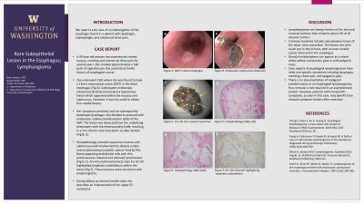
Case Description/Methods: A 49 year-old woman has experienced chronic nausea, vomiting and substernal chest pain for several years. She smoked approximately a half pack of cigarettes per day and had no family history of esophageal cancer. She underwent EGD where she was found to have a 12mm small submucosal tumor (SMT) in the distal esophagus (Fig A). Endoscopic biopsy was obtained but non-diagnostic. Subsequent endoscopic ultrasound (EUS) demonstrated a hypoechoic lesion which appeared within mucosa and submucosa. However, it was too small to obtain fine-needle biopsy. Her symptoms persisted and she subsequently developed dysphagia. After discussion with the therapeutic endoscopist, she decided to proceed with endoscopic submucosal dissection (ESD) of the SMT. The lesion was dissected from the underlying deep layers with the electrocautery knife, resulting in a 14 x 20 mm area resected in en-bloc fashion (Fig B, C). Histopathology revealed squamous mucosa and submucosa with involvement by dilated, ectatic and anastomosing lymphatic spaces lined by flat, bland appearing endothelial cells with thin proteinaceous material and admixed lymphocytes (Fig D, E). An immunohistochemical stain for D2-40 highlighted lymphatic endothelium within the lesion (Fig F). The histomorphologic features were consistent with lymphangioma. During follow-up several months later, she describes an improvement of her upper GI symptoms.
Discussion: Lymphangiomas are benign lesions of the skin and mucosal surfaces that comprise about 4% of all vascular tumors. Common locations include subcutaneous tissue of the head, neck and axillae. The lesions are seen much less in the GI tract, with an even smaller subset observed in the esophagus. Grossly lymphangioma can appear as a raised white-yellow, translucent, grey or pink polypoid mass. Case reports of esophageal lymphangiomas have cited non-specific symptoms including dysphagia, vomiting, chest pain, and epigastric pain. There is no documentation of malignant transformation in an esophageal lymphangioma, thus removal is not required in an asymptomatic patient. However, patients with nonspecific symptoms, as seen in this case, may have benefit from reduced symptom burden after resection.

Disclosures:
Neha Naidoo, MD, Deepti Reddi, MD, Yutaka Tomizawa, MD. P1917 - Rare Subepithelial Lesion in the Esophagus: Lymphangioma, ACG 2023 Annual Scientific Meeting Abstracts. Vancouver, BC, Canada: American College of Gastroenterology.
University of Washington, Seattle, WA
Introduction: Lymphangiomas of the gastrointestinal (GI) tract are uncommon, and are more frequently found in the head, neck, and axilla. We report a rare case of a lymphangioma of the esophagus found in a patient with dysphagia, odynophagia, and substernal chest pain.
Case Description/Methods: A 49 year-old woman has experienced chronic nausea, vomiting and substernal chest pain for several years. She smoked approximately a half pack of cigarettes per day and had no family history of esophageal cancer. She underwent EGD where she was found to have a 12mm small submucosal tumor (SMT) in the distal esophagus (Fig A). Endoscopic biopsy was obtained but non-diagnostic. Subsequent endoscopic ultrasound (EUS) demonstrated a hypoechoic lesion which appeared within mucosa and submucosa. However, it was too small to obtain fine-needle biopsy. Her symptoms persisted and she subsequently developed dysphagia. After discussion with the therapeutic endoscopist, she decided to proceed with endoscopic submucosal dissection (ESD) of the SMT. The lesion was dissected from the underlying deep layers with the electrocautery knife, resulting in a 14 x 20 mm area resected in en-bloc fashion (Fig B, C). Histopathology revealed squamous mucosa and submucosa with involvement by dilated, ectatic and anastomosing lymphatic spaces lined by flat, bland appearing endothelial cells with thin proteinaceous material and admixed lymphocytes (Fig D, E). An immunohistochemical stain for D2-40 highlighted lymphatic endothelium within the lesion (Fig F). The histomorphologic features were consistent with lymphangioma. During follow-up several months later, she describes an improvement of her upper GI symptoms.
Discussion: Lymphangiomas are benign lesions of the skin and mucosal surfaces that comprise about 4% of all vascular tumors. Common locations include subcutaneous tissue of the head, neck and axillae. The lesions are seen much less in the GI tract, with an even smaller subset observed in the esophagus. Grossly lymphangioma can appear as a raised white-yellow, translucent, grey or pink polypoid mass. Case reports of esophageal lymphangiomas have cited non-specific symptoms including dysphagia, vomiting, chest pain, and epigastric pain. There is no documentation of malignant transformation in an esophageal lymphangioma, thus removal is not required in an asymptomatic patient. However, patients with nonspecific symptoms, as seen in this case, may have benefit from reduced symptom burden after resection.

Figure: A. a SMT in the distal esophagus, B. C. an area resected by ESD in en-bloc fashion, D. 20x lower power field, E. x100 high power field, F. D2-40 IHC stain 100x
Disclosures:
Neha Naidoo indicated no relevant financial relationships.
Deepti Reddi indicated no relevant financial relationships.
Yutaka Tomizawa: Boston Scientific Company – Consultant. Medtronic – Consultant.
Neha Naidoo, MD, Deepti Reddi, MD, Yutaka Tomizawa, MD. P1917 - Rare Subepithelial Lesion in the Esophagus: Lymphangioma, ACG 2023 Annual Scientific Meeting Abstracts. Vancouver, BC, Canada: American College of Gastroenterology.

