Monday Poster Session
Category: Esophagus
P1928 - Wrapped Around: Bariatric Surgery Related Adhesion Causing Pseudoachalasia
Monday, October 23, 2023
10:30 AM - 4:15 PM PT
Location: Exhibit Hall

Has Audio

Thanita Thongtan, MD
University of Texas Rio Grande Valley
Atlanta, GA
Presenting Author(s)
Thanita Thongtan, MD1, Carlos Cardenas, MD2, Murthy Badiga, MD, FACG3, Asif Zamir, MD, FACG3
1University of Texas Rio Grande Valley, Edinburg, TX; 2DHR Health, Edinburg, TX; 3DHR Health Gastroenterology, Edinburg, TX
Introduction: Pseudoachalasia, accounts for just 4% of achalasia cases. It typically arises as a secondary esophageal dysfunction, often linked to malignancy. Pseudoachalasia stemming from bariatric post-operative complications is exceedingly rare. Here, we present a unique case of pseudoachalasia caused by late complications from bariatric surgery, specifically adhesions.
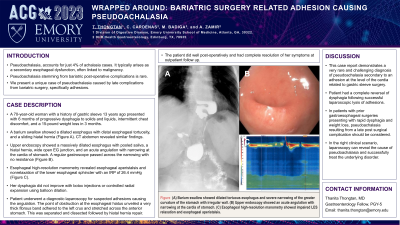
Case Description/Methods: A 70-year-old woman with a history of gastric sleeve 13 years ago presented with 6 months of progressive dysphagia to solids and liquids, intermittent chest discomfort, and a 15-pound weight loss in 3 months. A barium swallow showed a dilated esophagus with distal esophageal tortuosity, and a sliding hiatal hernia (Figure 1). CT abdomen revealed similar findings. Upper endoscopy showed a massively dilated esophagus with pooled saliva, a hiatal hernia, wide open EG junction, and an acute angulation with narrowing at the cardia of stomach. A regular gastroscope passed across the narrowing with no resistance (Figure 2). Esophageal high-resolution manometry revealed esophageal aperistalsis and nonrelaxation of the lower esophageal sphincter with an IRP of 26.4 mmHg (Figure 3). Her dysphagia did not improve with botox injections or controlled radial expansion using balloon dilation. Patient underwent a diagnostic laparoscopy for suspected adhesions causing the angulation. The point of obstruction at the esophageal hiatus unveiled a very thick fibrous band adhered to the left crus and stretched across the anterior stomach. This was separated and dissected followed by hiatal hernia repair. The patient did well post-operatively and had complete resolution of her symptoms at outpatient follow up.
Discussion: This case report demonstrates a very rare and challenging diagnosis of pseudoachalasia secondary to an adhesion at the level of the cardia related to gastric sleeve surgery. Patient had a complete reversal of dysphagia following successful laparoscopic lysis of adhesions. In patients with prior gastroesophageal surgeries presenting with rapid dysphagia and weight loss, pseudoachalasia resulting from a late post surgical complication should be considered. In the right clinical scenario, laparoscopy can reveal the cause of pseudoachalasia and successfully treat the underlying disorder.

Disclosures:
Thanita Thongtan, MD1, Carlos Cardenas, MD2, Murthy Badiga, MD, FACG3, Asif Zamir, MD, FACG3. P1928 - Wrapped Around: Bariatric Surgery Related Adhesion Causing Pseudoachalasia, ACG 2023 Annual Scientific Meeting Abstracts. Vancouver, BC, Canada: American College of Gastroenterology.
1University of Texas Rio Grande Valley, Edinburg, TX; 2DHR Health, Edinburg, TX; 3DHR Health Gastroenterology, Edinburg, TX
Introduction: Pseudoachalasia, accounts for just 4% of achalasia cases. It typically arises as a secondary esophageal dysfunction, often linked to malignancy. Pseudoachalasia stemming from bariatric post-operative complications is exceedingly rare. Here, we present a unique case of pseudoachalasia caused by late complications from bariatric surgery, specifically adhesions.
Case Description/Methods: A 70-year-old woman with a history of gastric sleeve 13 years ago presented with 6 months of progressive dysphagia to solids and liquids, intermittent chest discomfort, and a 15-pound weight loss in 3 months. A barium swallow showed a dilated esophagus with distal esophageal tortuosity, and a sliding hiatal hernia (Figure 1). CT abdomen revealed similar findings. Upper endoscopy showed a massively dilated esophagus with pooled saliva, a hiatal hernia, wide open EG junction, and an acute angulation with narrowing at the cardia of stomach. A regular gastroscope passed across the narrowing with no resistance (Figure 2). Esophageal high-resolution manometry revealed esophageal aperistalsis and nonrelaxation of the lower esophageal sphincter with an IRP of 26.4 mmHg (Figure 3). Her dysphagia did not improve with botox injections or controlled radial expansion using balloon dilation. Patient underwent a diagnostic laparoscopy for suspected adhesions causing the angulation. The point of obstruction at the esophageal hiatus unveiled a very thick fibrous band adhered to the left crus and stretched across the anterior stomach. This was separated and dissected followed by hiatal hernia repair. The patient did well post-operatively and had complete resolution of her symptoms at outpatient follow up.
Discussion: This case report demonstrates a very rare and challenging diagnosis of pseudoachalasia secondary to an adhesion at the level of the cardia related to gastric sleeve surgery. Patient had a complete reversal of dysphagia following successful laparoscopic lysis of adhesions. In patients with prior gastroesophageal surgeries presenting with rapid dysphagia and weight loss, pseudoachalasia resulting from a late post surgical complication should be considered. In the right clinical scenario, laparoscopy can reveal the cause of pseudoachalasia and successfully treat the underlying disorder.

Figure: Figure (A) Barium swallow showed dilated tortuous esophagus and severe narrowing of the greater curvature of the stomach with irregular wall. (B) Upper endoscopy showed an acute angulation with narrowing at the cardia of stomach. (C) Esophageal high-resolution manometry showed impaired LES relaxation and esophageal aperistalsis.
Disclosures:
Thanita Thongtan indicated no relevant financial relationships.
Carlos Cardenas indicated no relevant financial relationships.
Murthy Badiga indicated no relevant financial relationships.
Asif Zamir indicated no relevant financial relationships.
Thanita Thongtan, MD1, Carlos Cardenas, MD2, Murthy Badiga, MD, FACG3, Asif Zamir, MD, FACG3. P1928 - Wrapped Around: Bariatric Surgery Related Adhesion Causing Pseudoachalasia, ACG 2023 Annual Scientific Meeting Abstracts. Vancouver, BC, Canada: American College of Gastroenterology.
