Monday Poster Session
Category: Esophagus
P1934 - Endoscopic Retrieval of a Wireless pH Capsule Dropped Into the Lung
Monday, October 23, 2023
10:30 AM - 4:15 PM PT
Location: Exhibit Hall

Has Audio
- AL
Andy Lin, MD
University of California Irvine
Orange, California
Presenting Author(s)
Andy Lin, MD, Anastasia Chahine, MD, Amirali Tavangar, MD, Peter Nguyen, MD, Samuel Ji, MD, Khaldoon Khirfan, MD, Jason Samarasena, MD, John Lee, MD
University of California Irvine, Orange, CA
Introduction: Symptomatic gastroesophageal reflux disease (GERD) is very common in North America, with a 2014 study estimating prevalence at 18.1% to 27.8 %. Ambulatory pH monitoring is the preferred gold standard diagnostic modality, with the catheter-based esophageal monitoring being the most widely used test to correlate reflux with symptomology. Capsule-pH reflux testing has been introduced as a wireless system intended to replicate the traditional catheter-based monitoring without the drawbacks of patient discomfort. However, the nature of the this wireless system does lend itself to misplacement if direct guidance is not utilized. We present a case of capsule deployment in the oropharynx and subsequent aspiration.
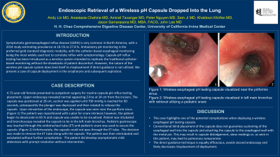
Case Description/Methods: A 72-year-old female presented to outpatient surgery for routine capsule-pH reflux testing placement. Upper endoscopy revealed normal appearing Z-line at 34 cm from the incisors. The capsule was positioned at 28 cm, suction was applied, and subsequently the plunger was depressed and then rotated to release the capsule. Upon reinsertion of the endoscope, the capsule was seen near the pyriform sinus. (Figure 1) The patient was repositioned with a plan for snare retrieval; however, the patient began to desaturate to 85 % and capsule was unable to be visualized. Patient was intubated and bronchoscopy revealed the capsule to be in the left main bronchus. Pediatric gastroscope was inserted through the endotracheal tube (ET) and pediatric snare was used to secure the capsule. (Figure 2) Unfortunately, the capsule could not pass through the ET tube. The decision was made to remove the ET tube along with the capsule. The patient was then reintubated and extubated without further complication. The patient did develop asymptomatic mild atelectasis with prompt resolution without intervention.
Discussion: This case highlights one of the potential complications when deploying a wireless esophageal pH testing capsule. Conventional blind placement of the capsule does not guarantee suctioning of the esophageal wall into the capsule and attaching the capsule to the esophageal wall with the metal pin. This may result in capsule dislodgement. This may skew readings or, as seen in this patient, may lead to pulmonary compromise. The direct guidance technique is equally efficacious, avoids second endoscopy and likely decreases misplacement of deployment.

Disclosures:
Andy Lin, MD, Anastasia Chahine, MD, Amirali Tavangar, MD, Peter Nguyen, MD, Samuel Ji, MD, Khaldoon Khirfan, MD, Jason Samarasena, MD, John Lee, MD. P1934 - Endoscopic Retrieval of a Wireless pH Capsule Dropped Into the Lung, ACG 2023 Annual Scientific Meeting Abstracts. Vancouver, BC, Canada: American College of Gastroenterology.
University of California Irvine, Orange, CA
Introduction: Symptomatic gastroesophageal reflux disease (GERD) is very common in North America, with a 2014 study estimating prevalence at 18.1% to 27.8 %. Ambulatory pH monitoring is the preferred gold standard diagnostic modality, with the catheter-based esophageal monitoring being the most widely used test to correlate reflux with symptomology. Capsule-pH reflux testing has been introduced as a wireless system intended to replicate the traditional catheter-based monitoring without the drawbacks of patient discomfort. However, the nature of the this wireless system does lend itself to misplacement if direct guidance is not utilized. We present a case of capsule deployment in the oropharynx and subsequent aspiration.
Case Description/Methods: A 72-year-old female presented to outpatient surgery for routine capsule-pH reflux testing placement. Upper endoscopy revealed normal appearing Z-line at 34 cm from the incisors. The capsule was positioned at 28 cm, suction was applied, and subsequently the plunger was depressed and then rotated to release the capsule. Upon reinsertion of the endoscope, the capsule was seen near the pyriform sinus. (Figure 1) The patient was repositioned with a plan for snare retrieval; however, the patient began to desaturate to 85 % and capsule was unable to be visualized. Patient was intubated and bronchoscopy revealed the capsule to be in the left main bronchus. Pediatric gastroscope was inserted through the endotracheal tube (ET) and pediatric snare was used to secure the capsule. (Figure 2) Unfortunately, the capsule could not pass through the ET tube. The decision was made to remove the ET tube along with the capsule. The patient was then reintubated and extubated without further complication. The patient did develop asymptomatic mild atelectasis with prompt resolution without intervention.
Discussion: This case highlights one of the potential complications when deploying a wireless esophageal pH testing capsule. Conventional blind placement of the capsule does not guarantee suctioning of the esophageal wall into the capsule and attaching the capsule to the esophageal wall with the metal pin. This may result in capsule dislodgement. This may skew readings or, as seen in this patient, may lead to pulmonary compromise. The direct guidance technique is equally efficacious, avoids second endoscopy and likely decreases misplacement of deployment.

Figure: Figure 1: Wireless esophageal pH testing capsule visualized near the piriformis sinus.
Figure 2: Wireless esophageal pH testing capsule visualized in left main bronchus with retrieval utilizing a pediatric snare
Figure 2: Wireless esophageal pH testing capsule visualized in left main bronchus with retrieval utilizing a pediatric snare
Disclosures:
Andy Lin indicated no relevant financial relationships.
Anastasia Chahine indicated no relevant financial relationships.
Amirali Tavangar indicated no relevant financial relationships.
Peter Nguyen indicated no relevant financial relationships.
Samuel Ji indicated no relevant financial relationships.
Khaldoon Khirfan indicated no relevant financial relationships.
Jason Samarasena: Applied Medical – Advisor or Review Panel Member. Boston Scientific – Consultant. Conmed – Consultant. Cook – Educational Grant. Neptune Medical – Consultant. Olympus – Consultant. Steris – Consultant.
John Lee indicated no relevant financial relationships.
Andy Lin, MD, Anastasia Chahine, MD, Amirali Tavangar, MD, Peter Nguyen, MD, Samuel Ji, MD, Khaldoon Khirfan, MD, Jason Samarasena, MD, John Lee, MD. P1934 - Endoscopic Retrieval of a Wireless pH Capsule Dropped Into the Lung, ACG 2023 Annual Scientific Meeting Abstracts. Vancouver, BC, Canada: American College of Gastroenterology.
