Monday Poster Session
Category: General Endoscopy
P2001 - Endoscopic Balloon Dilation of the Gastroesophageal Junction via Retroflexion for Progressive Achalasia
Monday, October 23, 2023
10:30 AM - 4:15 PM PT
Location: Exhibit Hall

Has Audio
- ZL
Zilan Lin, MD
Westchester Medical Center
Valhalla, NY
Presenting Author(s)
Zilan Lin, MD, Makeda Dawkins, MD, Virendra Tewari, MD
Westchester Medical Center, Valhalla, NY
Introduction: Pneumatic balloon dilation is an effective endoscopic modality for treating achalasia. Through the scope endoscopic balloon dilation is limited to a maximum of 20mm in diameter via forward view while effective treatment of achalasia requires larger diameter balloons of 30mm - 40mm. These are deployed under fluoroscopy which is often not feasible during routine endoscopy. We present a case of successful endoscopic balloon dilation of the gastroesophageal junction (GEJ) to 30mm in a patient with known history of achalasia by using a 20mm through the scope balloon with the endoscope in retroflexed position in the stomach.
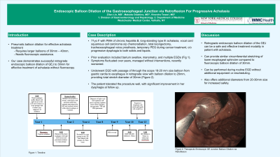
Case Description/Methods: A 71-year-old female with a history of chronic hepatitis B, long-standing type III achalasia, and vocal cord squamous cell carcinoma status post chemoradiation, total laryngectomy, tracheoesophageal voice prosthesis, and temporary percutaneous endoscopic gastrostomy during cancer treatment presented with recurrent, progressive dysphagia to both solids and liquids. Prior evaluation included barium swallow, manometry, and multiple esophagogastroduodenoscopies (EGDs) (Figure 1). Her symptoms fluctuated over years, and she managed her problem without specific interventions. Eventually she underwent another EGD due to progressive dysphagia. A through the scope 18-20 mm balloon was passed from the stomach into the esophagus via the retroflexed scope view. The balloon was then dilated to 20mm, resulting in the total dilatation to 30mm (Figure 2). The patient tolerated the procedure without complication, and she reported significant improvement in her dysphagia at follow up.
Discussion: Retrograde endoscopic balloon dilation of the GEJ was a safe and effective treatment modality in this case of achalasia, providing similar circumferential stretching of lower esophageal sphincter compared to fluoroscopic balloon dilation of 30mm. Although this method leaves a different GEJ shape compared to fluoroscopy balloon deployment, inflation under direct visualization has an added safety profile with a lower rate of esophageal perforation by providing an additional diameter range between 20-30mm. In addition, treatment can be offered during routine EGD in patients with achalasia without requiring additional equipment or rescheduling.

Disclosures:
Zilan Lin, MD, Makeda Dawkins, MD, Virendra Tewari, MD. P2001 - Endoscopic Balloon Dilation of the Gastroesophageal Junction via Retroflexion for Progressive Achalasia, ACG 2023 Annual Scientific Meeting Abstracts. Vancouver, BC, Canada: American College of Gastroenterology.
Westchester Medical Center, Valhalla, NY
Introduction: Pneumatic balloon dilation is an effective endoscopic modality for treating achalasia. Through the scope endoscopic balloon dilation is limited to a maximum of 20mm in diameter via forward view while effective treatment of achalasia requires larger diameter balloons of 30mm - 40mm. These are deployed under fluoroscopy which is often not feasible during routine endoscopy. We present a case of successful endoscopic balloon dilation of the gastroesophageal junction (GEJ) to 30mm in a patient with known history of achalasia by using a 20mm through the scope balloon with the endoscope in retroflexed position in the stomach.
Case Description/Methods: A 71-year-old female with a history of chronic hepatitis B, long-standing type III achalasia, and vocal cord squamous cell carcinoma status post chemoradiation, total laryngectomy, tracheoesophageal voice prosthesis, and temporary percutaneous endoscopic gastrostomy during cancer treatment presented with recurrent, progressive dysphagia to both solids and liquids. Prior evaluation included barium swallow, manometry, and multiple esophagogastroduodenoscopies (EGDs) (Figure 1). Her symptoms fluctuated over years, and she managed her problem without specific interventions. Eventually she underwent another EGD due to progressive dysphagia. A through the scope 18-20 mm balloon was passed from the stomach into the esophagus via the retroflexed scope view. The balloon was then dilated to 20mm, resulting in the total dilatation to 30mm (Figure 2). The patient tolerated the procedure without complication, and she reported significant improvement in her dysphagia at follow up.
Discussion: Retrograde endoscopic balloon dilation of the GEJ was a safe and effective treatment modality in this case of achalasia, providing similar circumferential stretching of lower esophageal sphincter compared to fluoroscopic balloon dilation of 30mm. Although this method leaves a different GEJ shape compared to fluoroscopy balloon deployment, inflation under direct visualization has an added safety profile with a lower rate of esophageal perforation by providing an additional diameter range between 20-30mm. In addition, treatment can be offered during routine EGD in patients with achalasia without requiring additional equipment or rescheduling.

Figure: Figure 1. Timeline. Figure 2. Therapeutic Endoscopic Gastroesophageal Junction Balloon Dilation via Retroflexion.
Disclosures:
Zilan Lin indicated no relevant financial relationships.
Makeda Dawkins indicated no relevant financial relationships.
Virendra Tewari indicated no relevant financial relationships.
Zilan Lin, MD, Makeda Dawkins, MD, Virendra Tewari, MD. P2001 - Endoscopic Balloon Dilation of the Gastroesophageal Junction via Retroflexion for Progressive Achalasia, ACG 2023 Annual Scientific Meeting Abstracts. Vancouver, BC, Canada: American College of Gastroenterology.
