Monday Poster Session
Category: General Endoscopy
P2010 - Cytomegalovirus Gastritis Mimicking a Gastroesophageal Junction Malignancy in an AIDS Patient
Monday, October 23, 2023
10:30 AM - 4:15 PM PT
Location: Exhibit Hall

Has Audio

Connor Lovingood, MD
University of Tennessee HSC College of Medicine Chattanooga
Chattanooga, TN
Presenting Author(s)
Connor Lovingood, MD, Nancy Jhanji, DO, James Pitcher, MD, William K. Oelsner, MD, Laxmi Parsa, MD
University of Tennessee HSC College of Medicine Chattanooga, Chattanooga, TN
Introduction: Cytomegalovirus (CMV) commonly undergoes reactivation in the setting of immunosuppression leading to a wide range of clinical manifestations. The most common CMV gastrointestinal manifestation is colitis. CMV gastritis has also been recorded in the literature, although much more infrequent. This case discusses a newly diagnosed HIV/AIDS patient found to have a malignant appearing GE Junction ulceration due to CMV in the absence of esophagitis or underlying malignancy.
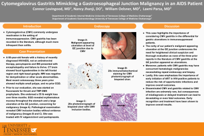
Case Description/Methods: A 60-year-old female with a history of recently diagnosed HIV/AIDS with CD4 count of 34, not on antiretroviral therapy (ART), pancytopenia and IBS presented with acute encephalopathy and failure to thrive. CT brain showed focal hypodensities in the left frontal region and right basal ganglia. MRI was negative for demyelination or other acute abnormalities. She denied NSAIDs, anticoagulation, antiplatelets, alcohol, smoking, or illicit drug use. Most recent colonoscopy was a few years prior with multiple small polyps, and no prior EGD. Prior to our evaluation, she was started on fluconazole for thrush and TMP-SMX prophylaxis. Gastroenterology was consulted for further evaluation of failure to thrive and dysphagia. She had a 55-pound weight loss over 2-3 months. EGD revealed erythematous mucosa throughout the stomach and a large ulceration at the GE junction, concerning for malignancy (Image A). Pathological examination revealed CMV inclusion bodies without evidence of malignancy (Images B and C). She was started on IV valganciclovir and pantoprazole. She represented two weeks later for failure to thrive. CT revealed numerous lytic lesions concerning for disseminated infection versus metastatic disease. Further workup was declined as she elected hospice care at this time.
Discussion: This case highlights the importance of considering CMV gastritis in the differential for gastric ulcerations in immunosuppressed patients. The rarity of our patient’s malignant appearing ulceration at the GE junction underscores the need for heightened clinical suspicion and thorough evaluation as none of the three prior reports in the literature of CMV gastritis at the GE junction appeared as ulcerations. Moreover, patients with CMV gastritis may have concurrent hematologic malignancies necessitating repeat endoscopy and biopsy. Lastly, this case emphasizes the importance of early initiation of ART in HIV-positive patients to reduce the risk of opportunistic infections and improve overall outcomes.

Disclosures:
Connor Lovingood, MD, Nancy Jhanji, DO, James Pitcher, MD, William K. Oelsner, MD, Laxmi Parsa, MD. P2010 - Cytomegalovirus Gastritis Mimicking a Gastroesophageal Junction Malignancy in an AIDS Patient, ACG 2023 Annual Scientific Meeting Abstracts. Vancouver, BC, Canada: American College of Gastroenterology.
University of Tennessee HSC College of Medicine Chattanooga, Chattanooga, TN
Introduction: Cytomegalovirus (CMV) commonly undergoes reactivation in the setting of immunosuppression leading to a wide range of clinical manifestations. The most common CMV gastrointestinal manifestation is colitis. CMV gastritis has also been recorded in the literature, although much more infrequent. This case discusses a newly diagnosed HIV/AIDS patient found to have a malignant appearing GE Junction ulceration due to CMV in the absence of esophagitis or underlying malignancy.
Case Description/Methods: A 60-year-old female with a history of recently diagnosed HIV/AIDS with CD4 count of 34, not on antiretroviral therapy (ART), pancytopenia and IBS presented with acute encephalopathy and failure to thrive. CT brain showed focal hypodensities in the left frontal region and right basal ganglia. MRI was negative for demyelination or other acute abnormalities. She denied NSAIDs, anticoagulation, antiplatelets, alcohol, smoking, or illicit drug use. Most recent colonoscopy was a few years prior with multiple small polyps, and no prior EGD. Prior to our evaluation, she was started on fluconazole for thrush and TMP-SMX prophylaxis. Gastroenterology was consulted for further evaluation of failure to thrive and dysphagia. She had a 55-pound weight loss over 2-3 months. EGD revealed erythematous mucosa throughout the stomach and a large ulceration at the GE junction, concerning for malignancy (Image A). Pathological examination revealed CMV inclusion bodies without evidence of malignancy (Images B and C). She was started on IV valganciclovir and pantoprazole. She represented two weeks later for failure to thrive. CT revealed numerous lytic lesions concerning for disseminated infection versus metastatic disease. Further workup was declined as she elected hospice care at this time.
Discussion: This case highlights the importance of considering CMV gastritis in the differential for gastric ulcerations in immunosuppressed patients. The rarity of our patient’s malignant appearing ulceration at the GE junction underscores the need for heightened clinical suspicion and thorough evaluation as none of the three prior reports in the literature of CMV gastritis at the GE junction appeared as ulcerations. Moreover, patients with CMV gastritis may have concurrent hematologic malignancies necessitating repeat endoscopy and biopsy. Lastly, this case emphasizes the importance of early initiation of ART in HIV-positive patients to reduce the risk of opportunistic infections and improve overall outcomes.

Figure: Figure 1:
Image A: Malignant-appearing ulceration at level of GE junction. Image B: 20x immunohistochemical staining for CMV photomicrograph of biopsied ulcer. Image C: 20x photomicrograph of biopsied ulcer with CMV inclusion bodies.
Image A: Malignant-appearing ulceration at level of GE junction. Image B: 20x immunohistochemical staining for CMV photomicrograph of biopsied ulcer. Image C: 20x photomicrograph of biopsied ulcer with CMV inclusion bodies.
Disclosures:
Connor Lovingood indicated no relevant financial relationships.
Nancy Jhanji indicated no relevant financial relationships.
James Pitcher indicated no relevant financial relationships.
William Oelsner indicated no relevant financial relationships.
Laxmi Parsa indicated no relevant financial relationships.
Connor Lovingood, MD, Nancy Jhanji, DO, James Pitcher, MD, William K. Oelsner, MD, Laxmi Parsa, MD. P2010 - Cytomegalovirus Gastritis Mimicking a Gastroesophageal Junction Malignancy in an AIDS Patient, ACG 2023 Annual Scientific Meeting Abstracts. Vancouver, BC, Canada: American College of Gastroenterology.
