Monday Poster Session
Category: GI Bleeding
P2055 - Jejunal Diverticular Bleeding: A Rare and Challenging Diagnosis Made by Video Capsule Endoscopy
Monday, October 23, 2023
10:30 AM - 4:15 PM PT
Location: Exhibit Hall

Has Audio
- FC
Fernando Contreras, MD
Centro de Gastroenterologia Avanzada
Santo Domingo, Distrito Nacional, Dominican Republic
Presenting Author(s)
Carlota Cabrer, MD1, Patricia Yunen, MD2, Ruddy Cabrera, MD2, Jose Gonzalez, MD3, Angel Gomez, MD2, Pedro Baez, MD2, Fernando Contreras, MD3
1Centro de Diagnostico Medicina Avanzada y Telemedicina, Santo Domingo, Distrito Nacional, Dominican Republic; 2Centro de Diagnóstico Medicina Avanzada y Telemedicina, Santo Domingo, Distrito Nacional, Dominican Republic; 3Centro de Gastroenterologia Avanzada, Santo Domingo, Distrito Nacional, Dominican Republic
Introduction: Small bowel (SB) diverticula are usually asymptomatic and are primarily found in the duodenum. They are rarer in the jejunum and ileum but can be present in all three segments in 3% of patients. Few cases have been reported of hemorrhage due to jejunal diverticulosis. SB diverticulosis complicated with bleeding requires prompt diagnosis and a therapeutic approach. In hemodynamically stable patients, capsule endoscopy is a valuable resource to assess the SB and the cause of bleeding. An accurate interpretation by a skilled observer is essential.
Case Description/Methods: A 68-year-old male with a past medical history of hypertension presented with melena for the past three days. His skin was pale on physical examination, borderline low blood pressure and tachycardia were noted. The abdomen with slightly augmented bowel sounds in rate and intensity. Rectal examination revealed soft black stool. Lab work: Hb of 8.1 g/dL, Hct 24.6%, rest within normal range.
The patient was admitted to ICU. During this presentation, he received 8 U of packed RBC. EGD and colonoscopy depicted no distinct source of bleeding. However, residual blood and clots were seen coming from the ileocecal valve, suggesting small bowel origin. A capsule endoscopy was deployed, and 5 minutes after identification of the first duodenal image, it suddenly showed a transition point of a smooth area with the absence of the villous pattern and a blood vessel with an adherent clot without active bleeding. The same image remained during the rest of the study. At 2 hours of recording, fresh blood became evident, raising the suspicion of a proximal SB diverticular bleed. A CTA A/P demonstrated jejunal diverticulosis.
The patient underwent anterograde SBE. Proximal jejunal diverticula with fresh blood and clots were seen without identification of the exact source of the bleed. Since the patient’s Hb continued to drop, an angiography was performed. The active bleeding site couldn’t be identified; therefore, embolization was not possible.
Partial proximal SB resection was performed. Bleeding control was achieved, and the patient recovered well.
Discussion: In this case, an unusual proximal jejunal diverticular bleeding was interestingly diagnosed by VCE after negative radiographic, EGD, and colonoscopy studies. The correct interpretation of VCE, in conjunction with a multidisciplinary approach, was crucial for a successful outcome.

Disclosures:
Carlota Cabrer, MD1, Patricia Yunen, MD2, Ruddy Cabrera, MD2, Jose Gonzalez, MD3, Angel Gomez, MD2, Pedro Baez, MD2, Fernando Contreras, MD3. P2055 - Jejunal Diverticular Bleeding: A Rare and Challenging Diagnosis Made by Video Capsule Endoscopy, ACG 2023 Annual Scientific Meeting Abstracts. Vancouver, BC, Canada: American College of Gastroenterology.
1Centro de Diagnostico Medicina Avanzada y Telemedicina, Santo Domingo, Distrito Nacional, Dominican Republic; 2Centro de Diagnóstico Medicina Avanzada y Telemedicina, Santo Domingo, Distrito Nacional, Dominican Republic; 3Centro de Gastroenterologia Avanzada, Santo Domingo, Distrito Nacional, Dominican Republic
Introduction: Small bowel (SB) diverticula are usually asymptomatic and are primarily found in the duodenum. They are rarer in the jejunum and ileum but can be present in all three segments in 3% of patients. Few cases have been reported of hemorrhage due to jejunal diverticulosis. SB diverticulosis complicated with bleeding requires prompt diagnosis and a therapeutic approach. In hemodynamically stable patients, capsule endoscopy is a valuable resource to assess the SB and the cause of bleeding. An accurate interpretation by a skilled observer is essential.
Case Description/Methods: A 68-year-old male with a past medical history of hypertension presented with melena for the past three days. His skin was pale on physical examination, borderline low blood pressure and tachycardia were noted. The abdomen with slightly augmented bowel sounds in rate and intensity. Rectal examination revealed soft black stool. Lab work: Hb of 8.1 g/dL, Hct 24.6%, rest within normal range.
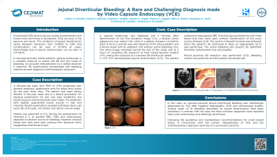
The patient was admitted to ICU. During this presentation, he received 8 U of packed RBC. EGD and colonoscopy depicted no distinct source of bleeding. However, residual blood and clots were seen coming from the ileocecal valve, suggesting small bowel origin. A capsule endoscopy was deployed, and 5 minutes after identification of the first duodenal image, it suddenly showed a transition point of a smooth area with the absence of the villous pattern and a blood vessel with an adherent clot without active bleeding. The same image remained during the rest of the study. At 2 hours of recording, fresh blood became evident, raising the suspicion of a proximal SB diverticular bleed. A CTA A/P demonstrated jejunal diverticulosis.
The patient underwent anterograde SBE. Proximal jejunal diverticula with fresh blood and clots were seen without identification of the exact source of the bleed. Since the patient’s Hb continued to drop, an angiography was performed. The active bleeding site couldn’t be identified; therefore, embolization was not possible.
Partial proximal SB resection was performed. Bleeding control was achieved, and the patient recovered well.
Discussion: In this case, an unusual proximal jejunal diverticular bleeding was interestingly diagnosed by VCE after negative radiographic, EGD, and colonoscopy studies. The correct interpretation of VCE, in conjunction with a multidisciplinary approach, was crucial for a successful outcome.

Figure: A) VCE showing smooth area with absence of the villous pattern and a blood vessel with an adherent clot without active bleeding.
B) The same area at 2 hours of recording with the presence of fresh blood and active bleeding.
C) CTA A/P demonstrating proximal jejunal diverticulosis.
D) Partial proximal SB resection surgical piece.
B) The same area at 2 hours of recording with the presence of fresh blood and active bleeding.
C) CTA A/P demonstrating proximal jejunal diverticulosis.
D) Partial proximal SB resection surgical piece.
Disclosures:
Carlota Cabrer indicated no relevant financial relationships.
Patricia Yunen indicated no relevant financial relationships.
Ruddy Cabrera indicated no relevant financial relationships.
Jose Gonzalez indicated no relevant financial relationships.
Angel Gomez indicated no relevant financial relationships.
Pedro Baez indicated no relevant financial relationships.
Fernando Contreras indicated no relevant financial relationships.
Carlota Cabrer, MD1, Patricia Yunen, MD2, Ruddy Cabrera, MD2, Jose Gonzalez, MD3, Angel Gomez, MD2, Pedro Baez, MD2, Fernando Contreras, MD3. P2055 - Jejunal Diverticular Bleeding: A Rare and Challenging Diagnosis Made by Video Capsule Endoscopy, ACG 2023 Annual Scientific Meeting Abstracts. Vancouver, BC, Canada: American College of Gastroenterology.
