Monday Poster Session
Category: GI Bleeding
P2076 - Meckel’s Diverticulum Diagnosed by CT Angiography
Monday, October 23, 2023
10:30 AM - 4:15 PM PT
Location: Exhibit Hall

Has Audio

Shamin H. Mahmud
Weill Cornell Medicine-Qatar
Doha, Ar Rayyan, Qatar
Presenting Author(s)
Shamin H. Mahmud, 1, David Wan, BS, MD2, Vladislav Fomin, MD3
1Weill Cornell Medicine-Qatar, Doha, Ar Rayyan, Qatar; 2Weill Cornell Medicine, New York, NY; 3New York-Presbyterian Hospital/Weill Cornell Medical College, New York, NY
Introduction: Meckel’s diverticulum (MD) is the most common gastrointestinal congenital malformation, arising from ectopic gastric mucosa. In adults, although uncommon and often asymptomatic, Meckel’s diverticulae can present with gastrointestinal bleeding.
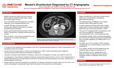
Case Description/Methods: A 19-year-old man presented to the emergency room with a syncopal episode preceded by 4 days of hematochezia, with associated nausea and vomiting. He was found to be tachycardic to 140 bpm and hypotensive to 109/57 mmHg, with physical exam notable for conjunctival pallor without abdominal tenderness or distension. Laboratory evaluation demonstrated iron-deficiency anemia with a hemoglobin of 4.9 g/dL, and a BUN:Cr ratio of 25. CT angiography of the abdomen revealed a diverticular outpouching measuring 2.5cm x 2.3cm x 0.7cm extending from a dilated small bowel loop in the right abdomen with active intraluminal contrast extravasation at its base, suspicious for a bleeding Meckel's diverticulum. Patient underwent exploratory laparotomy and resection of a Meckel’s diverticulum, located 100cm from the cecum. He was then discharged on postoperative day 2 after improvement of hemodynamics and hemoglobin.
Discussion: Meckel’s diverticulum is most often clinically silent in adults, with a low probability of developing gastrointestinal bleeding. The pathophysiology of bleeding is due to peptic ulceration within ectopic gastric mucosa. A nuclear medicine technetium-99m scan to detect ectopic gastric mucosa is typically used in pediatric patients but can have limited sensitivity in adults. The use of CT to diagnose MD is increasing, but is still rare due to the difficulty in distinguishing uncomplicated MD from a normal small bowel loop. Characteristic findings of active contrast extravasation into a diverticular outpouching arising from the terminal ileum are highly suggestive of a bleeding MD on a CT angiogram. The ability of a CT scan to detect a Meckel’s diverticulum is positively correlated with size, symptomatology, and amount of peritoneal fat. In our patient, the MD had active bleeding and was sufficiently large enough to be detected by CT. With increasing use of CT angiography for the diagnosis of lower GI bleeding we can expect an increased incidence of diagnosis of MD via this modality.
1. Elsayes, Khaled M., et al. "Imaging manifestations of Meckel's diverticulum." American Journal of Roentgenology 189.1 (2007): 81-88.

Disclosures:
Shamin H. Mahmud, 1, David Wan, BS, MD2, Vladislav Fomin, MD3. P2076 - Meckel’s Diverticulum Diagnosed by CT Angiography, ACG 2023 Annual Scientific Meeting Abstracts. Vancouver, BC, Canada: American College of Gastroenterology.
1Weill Cornell Medicine-Qatar, Doha, Ar Rayyan, Qatar; 2Weill Cornell Medicine, New York, NY; 3New York-Presbyterian Hospital/Weill Cornell Medical College, New York, NY
Introduction: Meckel’s diverticulum (MD) is the most common gastrointestinal congenital malformation, arising from ectopic gastric mucosa. In adults, although uncommon and often asymptomatic, Meckel’s diverticulae can present with gastrointestinal bleeding.
Case Description/Methods: A 19-year-old man presented to the emergency room with a syncopal episode preceded by 4 days of hematochezia, with associated nausea and vomiting. He was found to be tachycardic to 140 bpm and hypotensive to 109/57 mmHg, with physical exam notable for conjunctival pallor without abdominal tenderness or distension. Laboratory evaluation demonstrated iron-deficiency anemia with a hemoglobin of 4.9 g/dL, and a BUN:Cr ratio of 25. CT angiography of the abdomen revealed a diverticular outpouching measuring 2.5cm x 2.3cm x 0.7cm extending from a dilated small bowel loop in the right abdomen with active intraluminal contrast extravasation at its base, suspicious for a bleeding Meckel's diverticulum. Patient underwent exploratory laparotomy and resection of a Meckel’s diverticulum, located 100cm from the cecum. He was then discharged on postoperative day 2 after improvement of hemodynamics and hemoglobin.
Discussion: Meckel’s diverticulum is most often clinically silent in adults, with a low probability of developing gastrointestinal bleeding. The pathophysiology of bleeding is due to peptic ulceration within ectopic gastric mucosa. A nuclear medicine technetium-99m scan to detect ectopic gastric mucosa is typically used in pediatric patients but can have limited sensitivity in adults. The use of CT to diagnose MD is increasing, but is still rare due to the difficulty in distinguishing uncomplicated MD from a normal small bowel loop. Characteristic findings of active contrast extravasation into a diverticular outpouching arising from the terminal ileum are highly suggestive of a bleeding MD on a CT angiogram. The ability of a CT scan to detect a Meckel’s diverticulum is positively correlated with size, symptomatology, and amount of peritoneal fat. In our patient, the MD had active bleeding and was sufficiently large enough to be detected by CT. With increasing use of CT angiography for the diagnosis of lower GI bleeding we can expect an increased incidence of diagnosis of MD via this modality.
1. Elsayes, Khaled M., et al. "Imaging manifestations of Meckel's diverticulum." American Journal of Roentgenology 189.1 (2007): 81-88.

Figure: Figure 1: CT angiogram of the abdomen showing the Meckel’s Diverticulum (green arrow) as a diverticular outpouching from a dilated small bowel loop.
Disclosures:
Shamin Mahmud indicated no relevant financial relationships.
David Wan indicated no relevant financial relationships.
Vladislav Fomin indicated no relevant financial relationships.
Shamin H. Mahmud, 1, David Wan, BS, MD2, Vladislav Fomin, MD3. P2076 - Meckel’s Diverticulum Diagnosed by CT Angiography, ACG 2023 Annual Scientific Meeting Abstracts. Vancouver, BC, Canada: American College of Gastroenterology.
