Monday Poster Session
Category: GI Bleeding
P2091 - Not so Simple Exulceratio Simplex (Dieulafoy) – an Unusual Location for Massive GI Bleeding
Monday, October 23, 2023
10:30 AM - 4:15 PM PT
Location: Exhibit Hall

Has Audio

Anudeep S. Nakirikanti, BS
Emory University School of Medicine
Atlanta, Georgia
Presenting Author(s)
Anudeep S. Nakirikanti, BS1, Akshat Arya, MBBS2, Aunchalee Jaroenlapnopparat, MD3, Victoria Earl, MD1, Nicole Ruiz, MD4, Ibrahim Abubakar, MD4
1Emory University School of Medicine, Atlanta, GA; 2B.J. Medical College and Civil Hospital, Ahmedabad, Gujarat, India; 3Mount Auburn Hospital/Harvard Medical School, Cambridge, MA; 4Emory University, Atlanta, GA
Introduction: Dieulafoy lesions, consisting of submucosal arteries that erode into the overlying epithelium, are a rare source of upper GI bleeding that carry considerable morbidity and mortality. The lesion is predominantly found in the stomach with rare cases reported in the distal esophagus. Here, we describe a unique case of a large Dieulafoy lesion in the mid-esophagus.
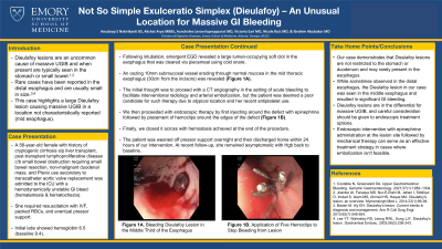
Case Description/Methods: A 56-year-old female with history of cryptogenic cirrhosis status post liver transplant, post-transplant lymphoproliferative disease status post small bowel resection, and non-malignant duodenal mass presented to the hospital with large volume hematochezia and hematemesis for one day. Exam showed tachycardia to 102 bpm, hypotension, pale conjunctiva, and bloody stools. The patient was on Plavix, last taken the prior afternoon. Labs revealed a hemoglobin of 6.5 (baseline from months before of 9.4) mg/dL. The patient was resuscitated in the emergency room and transferred to the ICU, where she continued to pass multiple bright red stools and ultimately required pressor support. The patient was intubated for airway protection, and emergent EGD showed large amounts of frank red blood and a lumen-occupying soft, red clot. The clot was cleared by careful piecemeal resection using 13mm cold snare. A 10 mm defect in the mucosa was seen at about 30 cm from the incisors with an apparent protruding submucosal vessel eroding through normal mucosa (figure 1A). The lesion appeared pulsatile with active oozing around the defect. Endoscopic therapy was pursued by injecting an 8 ml epinephrine solution in 1 ml aliquots around the lesion, followed by the application of ultra hemoclips beginning at the edge of the oozing vessel. In the end, hemoclips were deployed to close the defect and hemostasis was achieved (figure 1B). The patient was started on Pantoprazole IV drip and recovered with no further bleeding.
Discussion: Dieulafoy lesions in the esophagus are quite rare and when present are often located in the distal esophagus. To our knowledge, this is one of the few times a large Dieulafoy lesion was observed in the mid-esophagus. This was treated successfully with injection of epinephrine and through scope clips.

Disclosures:
Anudeep S. Nakirikanti, BS1, Akshat Arya, MBBS2, Aunchalee Jaroenlapnopparat, MD3, Victoria Earl, MD1, Nicole Ruiz, MD4, Ibrahim Abubakar, MD4. P2091 - Not so Simple Exulceratio Simplex (Dieulafoy) – an Unusual Location for Massive GI Bleeding, ACG 2023 Annual Scientific Meeting Abstracts. Vancouver, BC, Canada: American College of Gastroenterology.
1Emory University School of Medicine, Atlanta, GA; 2B.J. Medical College and Civil Hospital, Ahmedabad, Gujarat, India; 3Mount Auburn Hospital/Harvard Medical School, Cambridge, MA; 4Emory University, Atlanta, GA
Introduction: Dieulafoy lesions, consisting of submucosal arteries that erode into the overlying epithelium, are a rare source of upper GI bleeding that carry considerable morbidity and mortality. The lesion is predominantly found in the stomach with rare cases reported in the distal esophagus. Here, we describe a unique case of a large Dieulafoy lesion in the mid-esophagus.
Case Description/Methods: A 56-year-old female with history of cryptogenic cirrhosis status post liver transplant, post-transplant lymphoproliferative disease status post small bowel resection, and non-malignant duodenal mass presented to the hospital with large volume hematochezia and hematemesis for one day. Exam showed tachycardia to 102 bpm, hypotension, pale conjunctiva, and bloody stools. The patient was on Plavix, last taken the prior afternoon. Labs revealed a hemoglobin of 6.5 (baseline from months before of 9.4) mg/dL. The patient was resuscitated in the emergency room and transferred to the ICU, where she continued to pass multiple bright red stools and ultimately required pressor support. The patient was intubated for airway protection, and emergent EGD showed large amounts of frank red blood and a lumen-occupying soft, red clot. The clot was cleared by careful piecemeal resection using 13mm cold snare. A 10 mm defect in the mucosa was seen at about 30 cm from the incisors with an apparent protruding submucosal vessel eroding through normal mucosa (figure 1A). The lesion appeared pulsatile with active oozing around the defect. Endoscopic therapy was pursued by injecting an 8 ml epinephrine solution in 1 ml aliquots around the lesion, followed by the application of ultra hemoclips beginning at the edge of the oozing vessel. In the end, hemoclips were deployed to close the defect and hemostasis was achieved (figure 1B). The patient was started on Pantoprazole IV drip and recovered with no further bleeding.
Discussion: Dieulafoy lesions in the esophagus are quite rare and when present are often located in the distal esophagus. To our knowledge, this is one of the few times a large Dieulafoy lesion was observed in the mid-esophagus. This was treated successfully with injection of epinephrine and through scope clips.

Figure: Figure 1A) Bleeding Dieulafoy Lesion in the Middle Third of Esophagus, 1B) Five Hemoclips were placed around the Dieulafoy lesion to stop the bleeding.
Disclosures:
Anudeep Nakirikanti indicated no relevant financial relationships.
Akshat Arya indicated no relevant financial relationships.
Aunchalee Jaroenlapnopparat indicated no relevant financial relationships.
Victoria Earl indicated no relevant financial relationships.
Nicole Ruiz indicated no relevant financial relationships.
Ibrahim Abubakar indicated no relevant financial relationships.
Anudeep S. Nakirikanti, BS1, Akshat Arya, MBBS2, Aunchalee Jaroenlapnopparat, MD3, Victoria Earl, MD1, Nicole Ruiz, MD4, Ibrahim Abubakar, MD4. P2091 - Not so Simple Exulceratio Simplex (Dieulafoy) – an Unusual Location for Massive GI Bleeding, ACG 2023 Annual Scientific Meeting Abstracts. Vancouver, BC, Canada: American College of Gastroenterology.
