Monday Poster Session
Category: GI Bleeding
P2094 - Aneurysms of Gastroepiploic Artery: A Rare Cause of Upper GI Bleeding
Monday, October 23, 2023
10:30 AM - 4:15 PM PT
Location: Exhibit Hall

Has Audio

Maram Alenzi, MD
St. Elizabeth's Medical Center
Boston, MA
Presenting Author(s)
Maram Alenzi, MD1, Iyiad AlAbdul-Razzak, MD1, Saleh Alghsoon, MD2, Ali Jon, MD1, Derek Frederickson, MD1
1St. Elizabeth's Medical Center, Boston, MA; 2Tufts Medical Center, Boston, MA
Introduction: Gastroepiploic artery aneurysms (GEAAs) are rare accounting for 3–4% of all visceral arteries' aneurysms. They are usually silent, but potentially fatal as they are associated with 90% risk of rupture and 70% mortality rate after rupture due to life-threatening hemorrhage. Hence, the general approach to GEAAs is early elective intervention to minimize the risk of rupture. Here we present a rare case of three in-line large-size aneurysms of the right gastroepiploic artery that was successfully treated by transcatheter coil embolization.
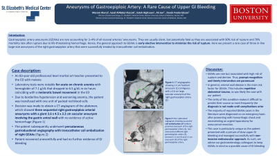
Case Description/Methods: An 83-year-old professional level martial art teacher presented to the ED with weakness, dyspnea, and melena. His laboratory tests were notable for acute on chronic anemia with hemoglobin of 7.1 g/dL that dropped to 6.5 g/dL in six hours coinciding with a melanotic bowel movement in the ED. A computed tomography (CT) of abdomen and pelvis with contrast was obtained revealing a large right gastroepiploic artery aneurysm measuring 3.5 cm. Due to borderline hypotension and worsening anemia, the patient was transfused with one unit of packed red blood cells. Decision was made to obtain a CT angiogram of the abdomen, which showed three sequential right gastroepiploic arterial aneurysms with a giant 3.6 x 4.5 x 3.2 cm saccular aneurysm involving the gastric antral wall with no evidence of active hemorrhage. The patient subsequently underwent percutaneous gastroduodenal angiography with transcatheter coil embolization of right GEAAs (Figure). He recovered uneventfully and had no further evidence of GI bleeding.
Discussion: GEAAs are rare but associated with high risk of rupture and demise. Thus, prompt recognition and timely intervention are paramount. In general, arterial wall disease is the main risk factor for GEAAs. This includes repetitive abdominal trauma, as was likely the case with our patient. The rarity of this condition makes it difficult to predict their course as most frequently the diagnosis is not made until complications arise. The majority of reported cases of GEAAs in the literature were diagnosed on an emergency basis after presenting with hemorrhagic shock and necessitating an urgent laparotomy for aneurysmal resection. This case is particularly unique as the patient presented with a picture of slow upper GI bleeding and managed successfully with non-invasive endovascular approach. As such, we advise our gastroenterology colleagues to keep GEAAs is mind as a possible cause of GI bleeding.

Disclosures:
Maram Alenzi, MD1, Iyiad AlAbdul-Razzak, MD1, Saleh Alghsoon, MD2, Ali Jon, MD1, Derek Frederickson, MD1. P2094 - Aneurysms of Gastroepiploic Artery: A Rare Cause of Upper GI Bleeding, ACG 2023 Annual Scientific Meeting Abstracts. Vancouver, BC, Canada: American College of Gastroenterology.
1St. Elizabeth's Medical Center, Boston, MA; 2Tufts Medical Center, Boston, MA
Introduction: Gastroepiploic artery aneurysms (GEAAs) are rare accounting for 3–4% of all visceral arteries' aneurysms. They are usually silent, but potentially fatal as they are associated with 90% risk of rupture and 70% mortality rate after rupture due to life-threatening hemorrhage. Hence, the general approach to GEAAs is early elective intervention to minimize the risk of rupture. Here we present a rare case of three in-line large-size aneurysms of the right gastroepiploic artery that was successfully treated by transcatheter coil embolization.
Case Description/Methods: An 83-year-old professional level martial art teacher presented to the ED with weakness, dyspnea, and melena. His laboratory tests were notable for acute on chronic anemia with hemoglobin of 7.1 g/dL that dropped to 6.5 g/dL in six hours coinciding with a melanotic bowel movement in the ED. A computed tomography (CT) of abdomen and pelvis with contrast was obtained revealing a large right gastroepiploic artery aneurysm measuring 3.5 cm. Due to borderline hypotension and worsening anemia, the patient was transfused with one unit of packed red blood cells. Decision was made to obtain a CT angiogram of the abdomen, which showed three sequential right gastroepiploic arterial aneurysms with a giant 3.6 x 4.5 x 3.2 cm saccular aneurysm involving the gastric antral wall with no evidence of active hemorrhage. The patient subsequently underwent percutaneous gastroduodenal angiography with transcatheter coil embolization of right GEAAs (Figure). He recovered uneventfully and had no further evidence of GI bleeding.
Discussion: GEAAs are rare but associated with high risk of rupture and demise. Thus, prompt recognition and timely intervention are paramount. In general, arterial wall disease is the main risk factor for GEAAs. This includes repetitive abdominal trauma, as was likely the case with our patient. The rarity of this condition makes it difficult to predict their course as most frequently the diagnosis is not made until complications arise. The majority of reported cases of GEAAs in the literature were diagnosed on an emergency basis after presenting with hemorrhagic shock and necessitating an urgent laparotomy for aneurysmal resection. This case is particularly unique as the patient presented with a picture of slow upper GI bleeding and managed successfully with non-invasive endovascular approach. As such, we advise our gastroenterology colleagues to keep GEAAs is mind as a possible cause of GI bleeding.

Figure: Figure 1: (A) CT angiography showing 1.5 cm fusiform aneurysm (1) contiguous with a 3.6 cm large saccular aneurysm of the right gastroepiploic artery (2). (B) Non subtracted angiogram showing fusiform proximal aneurysm of the right gastroepiploic artery (1), non-aneurysmal efferent right gastroepiploic artery (2) have been occluded with coil embolization. Coil embolization of terminal 1.5 cm (3) and 3.1 cm aneurysm (4) of the right gastroepiploic artery indicating sequential location of these aneurysms corresponding to the location on CT angiogram images.
Disclosures:
Maram Alenzi indicated no relevant financial relationships.
Iyiad AlAbdul-Razzak indicated no relevant financial relationships.
Saleh Alghsoon indicated no relevant financial relationships.
Ali Jon indicated no relevant financial relationships.
Derek Frederickson indicated no relevant financial relationships.
Maram Alenzi, MD1, Iyiad AlAbdul-Razzak, MD1, Saleh Alghsoon, MD2, Ali Jon, MD1, Derek Frederickson, MD1. P2094 - Aneurysms of Gastroepiploic Artery: A Rare Cause of Upper GI Bleeding, ACG 2023 Annual Scientific Meeting Abstracts. Vancouver, BC, Canada: American College of Gastroenterology.
