Monday Poster Session
Category: Interventional Endoscopy
P2331 - Poorly Differentiated Squamous and Glandular Carcinoma Arising from a Duplication Cyst
Monday, October 23, 2023
10:30 AM - 4:15 PM PT
Location: Exhibit Hall

Has Audio
- AA
Adnan Ansari, DO
Franciscan Health
Olympia Fields, IL
Presenting Author(s)
Adnan Ansari, DO, Mohammad Arfeen, DO, Zohair Ahmed, MD
Franciscan Health, Olympia Fields, IL
Introduction: Gastric duplication cysts are generally benign congenital anomalies; there have been rare instances reported in the literature of adenocarcinoma arising from within a duplication cyst. We present a case of poorly differentiated squamous and glandular carcinoma arising from a duplication cyst.
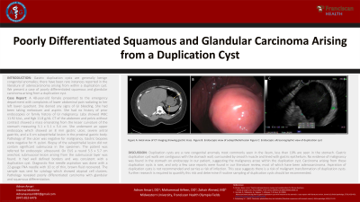
Case Description/Methods: A 48-year-old female presented to the emergency department with complaints of lower abdominal pain radiating to her left lower quadrant. She denied any signs of GI bleeding. She had been taking meloxicam and aspirin. She had no history of prior endoscopies or family history of GI malignancy. Labs showed WBC 11.92 k/uL, and Hgb 11.8 g/dL. CT of the abdomen and pelvis without contrast showed a mass emanating from the lesser curvature of the stomach measuring 5.1 x 5.1 x 5.4 cm. She underwent an upper endoscopy which showed an 8 mm gastric ulcer, severe antral gastritis, and a 6 cm subepithelial lesion in the proximal gastric body. Pathology of the ulcer was negative for malignancy. Gastric biopsies were negative for H. pylori. Biopsy of the subepithelial lesion did not contain significant submucosa in the specimen. The patient was referred for endoscopic ultrasound. On EUS a round 5.5 x 5.7 cm anechoic submucosal lesion arising from the submucosal layer was found. It had well defined borders and was consistent with a duplication cyst. Diagnostic fine needle aspiration was done with a 22-gauge FNA needle with 10 cc of thin, brown fluid recovered. The sample was sent for cytology which showed atypical cell clusters. Pathology revealed poorly differentiated carcinoma with glandular and squamous differentiation.
Discussion: Duplication cysts are a rare congenital anomaly most commonly seen in the ileum, less than 10% are seen in the stomach. Gastric duplication cyst walls are contiguous with the stomach wall, surrounded by smooth muscle and lined with gastric epithelium. No evidence of malignancy was found in the stomach on endoscopy in our patient, suggesting the malignancy arose within the duplication cyst. Carcinoma arising from these duplication cysts is rare, and only a few case reports were found in our literature review, most of which have been adenocarcinoma. Aspiration of duplication cysts is not recommended and carries a risk of infection. This case suggests there is a risk of malignant transformation of duplication cysts. Further research is required to quantify this risk and determine if routine sampling of duplication cysts should be recommended.

Disclosures:
Adnan Ansari, DO, Mohammad Arfeen, DO, Zohair Ahmed, MD. P2331 - Poorly Differentiated Squamous and Glandular Carcinoma Arising from a Duplication Cyst, ACG 2023 Annual Scientific Meeting Abstracts. Vancouver, BC, Canada: American College of Gastroenterology.
Franciscan Health, Olympia Fields, IL
Introduction: Gastric duplication cysts are generally benign congenital anomalies; there have been rare instances reported in the literature of adenocarcinoma arising from within a duplication cyst. We present a case of poorly differentiated squamous and glandular carcinoma arising from a duplication cyst.
Case Description/Methods: A 48-year-old female presented to the emergency department with complaints of lower abdominal pain radiating to her left lower quadrant. She denied any signs of GI bleeding. She had been taking meloxicam and aspirin. She had no history of prior endoscopies or family history of GI malignancy. Labs showed WBC 11.92 k/uL, and Hgb 11.8 g/dL. CT of the abdomen and pelvis without contrast showed a mass emanating from the lesser curvature of the stomach measuring 5.1 x 5.1 x 5.4 cm. She underwent an upper endoscopy which showed an 8 mm gastric ulcer, severe antral gastritis, and a 6 cm subepithelial lesion in the proximal gastric body. Pathology of the ulcer was negative for malignancy. Gastric biopsies were negative for H. pylori. Biopsy of the subepithelial lesion did not contain significant submucosa in the specimen. The patient was referred for endoscopic ultrasound. On EUS a round 5.5 x 5.7 cm anechoic submucosal lesion arising from the submucosal layer was found. It had well defined borders and was consistent with a duplication cyst. Diagnostic fine needle aspiration was done with a 22-gauge FNA needle with 10 cc of thin, brown fluid recovered. The sample was sent for cytology which showed atypical cell clusters. Pathology revealed poorly differentiated carcinoma with glandular and squamous differentiation.
Discussion: Duplication cysts are a rare congenital anomaly most commonly seen in the ileum, less than 10% are seen in the stomach. Gastric duplication cyst walls are contiguous with the stomach wall, surrounded by smooth muscle and lined with gastric epithelium. No evidence of malignancy was found in the stomach on endoscopy in our patient, suggesting the malignancy arose within the duplication cyst. Carcinoma arising from these duplication cysts is rare, and only a few case reports were found in our literature review, most of which have been adenocarcinoma. Aspiration of duplication cysts is not recommended and carries a risk of infection. This case suggests there is a risk of malignant transformation of duplication cysts. Further research is required to quantify this risk and determine if routine sampling of duplication cysts should be recommended.

Figure: Figure A: Axial view of CT imaging showing gastric mass.
Figure B: Endoscopic view of subepithelial lesion
Figure C: Endoscopic ultrasonographic view of duplication cyst
Figure B: Endoscopic view of subepithelial lesion
Figure C: Endoscopic ultrasonographic view of duplication cyst
Disclosures:
Adnan Ansari indicated no relevant financial relationships.
Mohammad Arfeen indicated no relevant financial relationships.
Zohair Ahmed indicated no relevant financial relationships.
Adnan Ansari, DO, Mohammad Arfeen, DO, Zohair Ahmed, MD. P2331 - Poorly Differentiated Squamous and Glandular Carcinoma Arising from a Duplication Cyst, ACG 2023 Annual Scientific Meeting Abstracts. Vancouver, BC, Canada: American College of Gastroenterology.
