Monday Poster Session
Category: Liver
P2459 - PEComa: A Rare Liver Tumor
Monday, October 23, 2023
10:30 AM - 4:15 PM PT
Location: Exhibit Hall

Has Audio

Christopher J. Costa, MD
University of Connecticut Health Center
Farmington, CT
Presenting Author(s)
Christopher J. Costa, MD1, Minh Thu T. Nguyen, MD2, Alexander Potashinksky, MD3
1University of Connecticut Health Center, Farmington, CT; 2UConn Health, Farmington, CT; 3Hospital of Central Connecticut, New Britain, CT
Introduction: Here we report a rare, benign hepatic tumor known as perivascular endothelial carcinoma (PEComa) with unique histologic and imaging findings. It should be considered in an expanded differential of a hepatic mass.
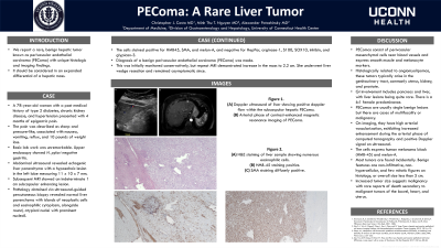
Case Description/Methods: A 78-year-old woman with a past medical history of type 2 diabetes, chronic kidney disease, and hypertension presented with 4 months of epigastric pain. The pain was described as sharp and pressure-like, associated with nausea, vomiting, reflux, and 10 pounds of weight loss. Basic labwork was unremarkable. Upper endoscopy showed H. pylori negative gastritis. Abdominal ultrasound revealed echogenic liver parenchyma with a hypoechoic lesion in the left lobe measuring 11 x 10 x 7 millimeters. Subsequent MRI showed an indeterminate 1 cm subcapsular enhancing lesion. Pathology obtained via ultrasound-guided percutaneous biopsy revealed normal liver parenchyma with islands of neoplastic cells and eosinophilic cytoplasm, alongside round, atypical nuclei with prominent nucleoli. The cells stained positive for HMB45, SMA, and melan-A, and negative for HepPar, arginase-1, S100, SOX10, inhibin, and glypican-3. Diagnosis of a benign perivascular endothelial carcinoma (PEComa) was made. This was initially monitored conservatively, but repeat MRI demonstrated increase in the mass to 2.2 cm. She underwent liver wedge resection.
Discussion: PEComas consist of perivascular mesenchymal cells located near blood vessels and express smooth muscle and melanocyte markers. Histologically related to angiomyolipomas, these tumors typically arise in the genitourinary tract, commonly uterus, kidney, and prostate. Gastrointestinal involvement includes pancreas and liver, with liver lesions being quite rare. There is a 6:1 female predominance. PEComas are usually single benign lesions but there are cases of multifocality or malignancy. On imaging, they have high arterial vascularization, exhibiting increased enhancement during the arterial phase of computed tomography and positive Doppler signal on ultrasound. Histologically, the cells express human melanoma black (HMB-45) and melan-A. Most tumors are found incidentally. Benign features are non-infiltrative, non-hypercellular, and few mitotic figures on histology, or overall size less than 5 cm. Increased tumor size suggests malignancy with rare reports of death secondary to malignant tumors of the bowel, heart, and uterus.

Disclosures:
Christopher J. Costa, MD1, Minh Thu T. Nguyen, MD2, Alexander Potashinksky, MD3. P2459 - PEComa: A Rare Liver Tumor, ACG 2023 Annual Scientific Meeting Abstracts. Vancouver, BC, Canada: American College of Gastroenterology.
1University of Connecticut Health Center, Farmington, CT; 2UConn Health, Farmington, CT; 3Hospital of Central Connecticut, New Britain, CT
Introduction: Here we report a rare, benign hepatic tumor known as perivascular endothelial carcinoma (PEComa) with unique histologic and imaging findings. It should be considered in an expanded differential of a hepatic mass.
Case Description/Methods: A 78-year-old woman with a past medical history of type 2 diabetes, chronic kidney disease, and hypertension presented with 4 months of epigastric pain. The pain was described as sharp and pressure-like, associated with nausea, vomiting, reflux, and 10 pounds of weight loss. Basic labwork was unremarkable. Upper endoscopy showed H. pylori negative gastritis. Abdominal ultrasound revealed echogenic liver parenchyma with a hypoechoic lesion in the left lobe measuring 11 x 10 x 7 millimeters. Subsequent MRI showed an indeterminate 1 cm subcapsular enhancing lesion. Pathology obtained via ultrasound-guided percutaneous biopsy revealed normal liver parenchyma with islands of neoplastic cells and eosinophilic cytoplasm, alongside round, atypical nuclei with prominent nucleoli. The cells stained positive for HMB45, SMA, and melan-A, and negative for HepPar, arginase-1, S100, SOX10, inhibin, and glypican-3. Diagnosis of a benign perivascular endothelial carcinoma (PEComa) was made. This was initially monitored conservatively, but repeat MRI demonstrated increase in the mass to 2.2 cm. She underwent liver wedge resection.
Discussion: PEComas consist of perivascular mesenchymal cells located near blood vessels and express smooth muscle and melanocyte markers. Histologically related to angiomyolipomas, these tumors typically arise in the genitourinary tract, commonly uterus, kidney, and prostate. Gastrointestinal involvement includes pancreas and liver, with liver lesions being quite rare. There is a 6:1 female predominance. PEComas are usually single benign lesions but there are cases of multifocality or malignancy. On imaging, they have high arterial vascularization, exhibiting increased enhancement during the arterial phase of computed tomography and positive Doppler signal on ultrasound. Histologically, the cells express human melanoma black (HMB-45) and melan-A. Most tumors are found incidentally. Benign features are non-infiltrative, non-hypercellular, and few mitotic figures on histology, or overall size less than 5 cm. Increased tumor size suggests malignancy with rare reports of death secondary to malignant tumors of the bowel, heart, and uterus.

Figure: Figure 1. (A) Doppler ultrasound of liver showing positive doppler flow within the subcaspular hepatic PECOMA. (B) Arterial phase of contrast enhanced magnetic resonance imaging of PECOMA
Disclosures:
Christopher Costa indicated no relevant financial relationships.
Minh Thu Nguyen indicated no relevant financial relationships.
Alexander Potashinksky indicated no relevant financial relationships.
Christopher J. Costa, MD1, Minh Thu T. Nguyen, MD2, Alexander Potashinksky, MD3. P2459 - PEComa: A Rare Liver Tumor, ACG 2023 Annual Scientific Meeting Abstracts. Vancouver, BC, Canada: American College of Gastroenterology.
