Monday Poster Session
Category: Liver
P2512 - Exploring Hepatic Consequences of Chronic MDMA Consumption: A Clinical Case Study and Histopathological Analysis
Monday, October 23, 2023
10:30 AM - 4:15 PM PT
Location: Exhibit Hall

Has Audio
.jpg)
Philip Bouchette, MD
LSU Health Sciences Center
Shreveport, LA
Presenting Author(s)
Philip Bouchette, MD, Simin Khan, MD, Maryam Mubashir, MD, Jacob Afude, MD, Jennifer Lee, MD, James Morris, MD
LSU Health Sciences Center, Shreveport, LA
Introduction: MDMA (3,4-methylenedioxymethamphetamine), also known as ecstasy or molly, is a synthetic amphetamine widely used as a recreational drug because it produces euphoria and increases sociability. Common side effects include nausea, muscle cramping, blurred vision, chills, & sweating. The liver is one of the most affected organs. We present a case of a 52-year-old woman with liver injury due to chronic MDMA use.
Case Description/Methods: A 52-year-old female with a past medical history of Osteoarthritis who presented with a chief complaint of diffuse abdominal pain. Patient reports the symptoms have been going on for about 2-3 weeks. She describes the pains as radiating to her flanks bilaterally, worse with movement, relieved with rest. She associates the pain with nausea/vomiting and poor oral intake. She also reports yellowing of her eyes for 1 week. Her urine had been darker than usual. Patient endorsed regular use of ecstasy (MDMA) over the past two years.
On her initial workup, her labs showed: aspartate aminotransferase AST of 1117/U/L, alanine aminotransferase ALT of 1866 U/L, Total bilirubin 1.0, INR 1.1, and lactate dehydrogenase of 422 U/L (Table 1). Gastroenterology was consulted for management recommendations. CT abdomen and pelvis showed no radiological evidence of an acute abdominopelvic abnormality. Interventional radiology was consulted for a percutaneous liver biopsy which was performed the subsequent hospital day.
After several days of supportive care, her liver function tests continued to improve with an AST of 681 and ALT of 1388, measured on the day of discharge. With resolution of symptoms, declining LFTs, she was deemed stable for discharge with referrals to follow up with GI outpatient. Pathology came back significant with necro- inflammatory patterns of liver injury, most likely resulting from MDMA drug insult (Figure 1).
Discussion:

Disclosures:
Philip Bouchette, MD, Simin Khan, MD, Maryam Mubashir, MD, Jacob Afude, MD, Jennifer Lee, MD, James Morris, MD. P2512 - Exploring Hepatic Consequences of Chronic MDMA Consumption: A Clinical Case Study and Histopathological Analysis, ACG 2023 Annual Scientific Meeting Abstracts. Vancouver, BC, Canada: American College of Gastroenterology.
LSU Health Sciences Center, Shreveport, LA
Introduction: MDMA (3,4-methylenedioxymethamphetamine), also known as ecstasy or molly, is a synthetic amphetamine widely used as a recreational drug because it produces euphoria and increases sociability. Common side effects include nausea, muscle cramping, blurred vision, chills, & sweating. The liver is one of the most affected organs. We present a case of a 52-year-old woman with liver injury due to chronic MDMA use.
Case Description/Methods: A 52-year-old female with a past medical history of Osteoarthritis who presented with a chief complaint of diffuse abdominal pain. Patient reports the symptoms have been going on for about 2-3 weeks. She describes the pains as radiating to her flanks bilaterally, worse with movement, relieved with rest. She associates the pain with nausea/vomiting and poor oral intake. She also reports yellowing of her eyes for 1 week. Her urine had been darker than usual. Patient endorsed regular use of ecstasy (MDMA) over the past two years.
On her initial workup, her labs showed: aspartate aminotransferase AST of 1117/U/L, alanine aminotransferase ALT of 1866 U/L, Total bilirubin 1.0, INR 1.1, and lactate dehydrogenase of 422 U/L (Table 1). Gastroenterology was consulted for management recommendations. CT abdomen and pelvis showed no radiological evidence of an acute abdominopelvic abnormality. Interventional radiology was consulted for a percutaneous liver biopsy which was performed the subsequent hospital day.
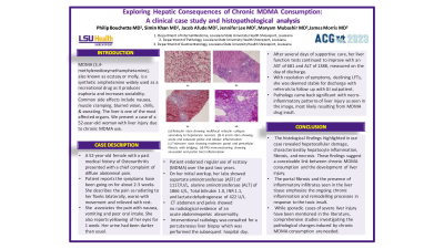
After several days of supportive care, her liver function tests continued to improve with an AST of 681 and ALT of 1388, measured on the day of discharge. With resolution of symptoms, declining LFTs, she was deemed stable for discharge with referrals to follow up with GI outpatient. Pathology came back significant with necro- inflammatory patterns of liver injury, most likely resulting from MDMA drug insult (Figure 1).
Discussion:
- The histological findings highlighted in our case revealed hepatocellular damage, characterized by hepatocyte inflammation, fibrosis, and necrosis. These findings suggest a conceivable link between chronic MDMA consumption and the development of liver injury
- The portal fibrosis and the presence of inflammatory infiltrates seen in the liver tissue emphasize the ongoing chronic inflammation and remodeling processes in response to the toxic insult
While sporadic cases of severe liver injury have been mentioned in the literature, comprehensive studies investigating the pathological changes induced by chronic MDMA consumption are needed

Figure: Figure 1. Pathology Findings: (a) Reticulin stain showing multifocal reticulin collapse secondary to hepatocyte necrosis. (b) H and E stain showing acute and subacute portal and lobular inflammation. (c) Trichrome stain showing moderate portal and pericellular fibrosis with bridging. (d) PAS immunostaining showing sinusoidal and portal tract inflammation.
Disclosures:
Philip Bouchette indicated no relevant financial relationships.
Simin Khan indicated no relevant financial relationships.
Maryam Mubashir indicated no relevant financial relationships.
Jacob Afude indicated no relevant financial relationships.
Jennifer Lee indicated no relevant financial relationships.
James Morris indicated no relevant financial relationships.
Philip Bouchette, MD, Simin Khan, MD, Maryam Mubashir, MD, Jacob Afude, MD, Jennifer Lee, MD, James Morris, MD. P2512 - Exploring Hepatic Consequences of Chronic MDMA Consumption: A Clinical Case Study and Histopathological Analysis, ACG 2023 Annual Scientific Meeting Abstracts. Vancouver, BC, Canada: American College of Gastroenterology.
