Monday Poster Session
Category: Liver
P2564 - Secretly Sarcoid
Monday, October 23, 2023
10:30 AM - 4:15 PM PT
Location: Exhibit Hall

Has Audio

Tricia A. Hengehold, MD
The Ohio State University
Columbus, OH
Presenting Author(s)
Tricia Hengehold, MD1, Chathur Acharya, MBBS2
1The Ohio State University, Columbus, OH; 2Ohio State University, Columbus, OH
Introduction: Sarcoidosis is a multisystem disease characterized by non-caseating granulomas. The pulmonary system is the most involved with parenchymal nodules and hilar lymphadenopathy. The liver is also often involved but can be missed as patients are usually asymptomatic. Hepatic sarcoid should be considered with chronic predominant cholestatic elevations in liver enzymes in the right clinical setting. Diagnosis should be confirmed by a liver biopsy as imaging is usually non-specific, though hepatomegaly is a common finding. We present a rare case of hepatic sarcoidosis that manifested as a liver mass on imaging.
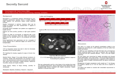
Case Description/Methods: A 57 yo F with stage IV low grade follicular lymphoma s/p bendamustine and rituximab 4 years prior, mild asymptomatic pulmonary sarcoidosis with possible hilar lymph node involvement, MS, and Hashimoto thyroiditis was seen in clinic for chronic elevated LFTs. Liver enzymes showed ALP had been persistently elevated in 100-200s for the past 5 years, intermittently elevated ALT with peak 138, and AST intermittently elevated with peak 66. Her abdominal CT scan showed heterogenous enhancing areas in segment 4/6 concerning for focal fatty sparing. Triple phase MRI with gadolinium (Fig A, arrow) showed a 1.7 x 1.5 cm T2 intermediate intense lesion with no washout in segment 6 that was concerning for a metastatic lesion/deposit such as lymphoma. Liver biopsy (Fig B) of the mass showed non-caseating granulomas, dense lymphocytic portal inflammation and bridging/nodular fibrosis consistent with hepatic sarcoidosis. Tissue from the rest of the liver showed simple hepatic steatosis and no fibrosis.
Discussion: This case highlights a rare radiological manifestation of sarcoidosis in the form of a homogeneous arterial enhancing lesion of the liver with no washout, no capsular enhancement and with restricted diffusion that can be easily mistaken for other solid malignancies of the liver. Although hepatic involvement is common in sarcoidosis, there are no characteristic radiological findings and nodular hepatic sarcoidosis has rarely been described. It was interesting to note in her biopsy that only the sarcoid nodule had intense inflammation and focal cirrhosis while the surrounding liver parenchyma biopsied did not have significant sarcoidosis or fibrosis. This indicates the patchy liver involvement in sarcoidosis and could explain the variation in natural history of hepatic sarcoidosis. It is important to be aware of this rare presentation in order to test and treat appropriately.

Disclosures:
Tricia Hengehold, MD1, Chathur Acharya, MBBS2. P2564 - Secretly Sarcoid, ACG 2023 Annual Scientific Meeting Abstracts. Vancouver, BC, Canada: American College of Gastroenterology.
1The Ohio State University, Columbus, OH; 2Ohio State University, Columbus, OH
Introduction: Sarcoidosis is a multisystem disease characterized by non-caseating granulomas. The pulmonary system is the most involved with parenchymal nodules and hilar lymphadenopathy. The liver is also often involved but can be missed as patients are usually asymptomatic. Hepatic sarcoid should be considered with chronic predominant cholestatic elevations in liver enzymes in the right clinical setting. Diagnosis should be confirmed by a liver biopsy as imaging is usually non-specific, though hepatomegaly is a common finding. We present a rare case of hepatic sarcoidosis that manifested as a liver mass on imaging.
Case Description/Methods: A 57 yo F with stage IV low grade follicular lymphoma s/p bendamustine and rituximab 4 years prior, mild asymptomatic pulmonary sarcoidosis with possible hilar lymph node involvement, MS, and Hashimoto thyroiditis was seen in clinic for chronic elevated LFTs. Liver enzymes showed ALP had been persistently elevated in 100-200s for the past 5 years, intermittently elevated ALT with peak 138, and AST intermittently elevated with peak 66. Her abdominal CT scan showed heterogenous enhancing areas in segment 4/6 concerning for focal fatty sparing. Triple phase MRI with gadolinium (Fig A, arrow) showed a 1.7 x 1.5 cm T2 intermediate intense lesion with no washout in segment 6 that was concerning for a metastatic lesion/deposit such as lymphoma. Liver biopsy (Fig B) of the mass showed non-caseating granulomas, dense lymphocytic portal inflammation and bridging/nodular fibrosis consistent with hepatic sarcoidosis. Tissue from the rest of the liver showed simple hepatic steatosis and no fibrosis.
Discussion: This case highlights a rare radiological manifestation of sarcoidosis in the form of a homogeneous arterial enhancing lesion of the liver with no washout, no capsular enhancement and with restricted diffusion that can be easily mistaken for other solid malignancies of the liver. Although hepatic involvement is common in sarcoidosis, there are no characteristic radiological findings and nodular hepatic sarcoidosis has rarely been described. It was interesting to note in her biopsy that only the sarcoid nodule had intense inflammation and focal cirrhosis while the surrounding liver parenchyma biopsied did not have significant sarcoidosis or fibrosis. This indicates the patchy liver involvement in sarcoidosis and could explain the variation in natural history of hepatic sarcoidosis. It is important to be aware of this rare presentation in order to test and treat appropriately.

Figure: Figure A with concerning lesion seen on MRI that prompted biopsy. Figure B with biopsy of the mass noting features suggestive of sarcoidosis such as dense lymphocytic portal inflammation, bridging/nodular fibrosis and and non-caseating granulomas.
Disclosures:
Tricia Hengehold indicated no relevant financial relationships.
Chathur Acharya indicated no relevant financial relationships.
Tricia Hengehold, MD1, Chathur Acharya, MBBS2. P2564 - Secretly Sarcoid, ACG 2023 Annual Scientific Meeting Abstracts. Vancouver, BC, Canada: American College of Gastroenterology.
