Monday Poster Session
Category: Liver
P2577 - Xanthogranulomatous Pyelonephritis: An Unusual Cause of Liver Abscesses
Monday, October 23, 2023
10:30 AM - 4:15 PM PT
Location: Exhibit Hall

Has Audio

Kelli Kosako Yost, MD
University of Arizona College of Medicine
Phoenix, AZ
Presenting Author(s)
Kelli Kosako Yost, MD, Dayna Telken, DO, Qumber Ali, BS, DO, Zaid Alkaissy, MD, Karn Wijarnpreecha, MD, MPH
University of Arizona College of Medicine, Phoenix, AZ
Introduction: Causes of hepatic abscesses include appendicitis, biliary tract disease, cholangitis, bile duct ischemia, and amoebic infections. Xanthogranulomatous pyelonephritis (XGP) is a rare sequela of chronic pyelonephritis involving the destruction of normal renal tissue and nonfunctioning granulomatous infiltration. It has the ability to extend into other solid organ structures, such as the liver or colon, causing extensive abscesses.
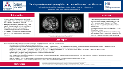
Case Description/Methods: The patient is a 68 year old male with history of treated hepatitis C, hypertension, benign prostatic hyperplasia, and diabetes, who presented with cough, shortness of breath with exertion, and fever. Upon admission, patient was found to be in septic shock with fever to 104.5 degrees F. CT abdomen/pelvis with contrast showed right inferior hepatic lobe lesions (4.9 cm x 3.1 cm and 5.3 cm x 4.5 cm) with peripheral enhancement, an enhancing exophytic lesion of the right kidney (4.4 cm x 3.9 cm) that was contiguous with hepatic lesions, extension of a sub-hepatic lesion adjacent to the right colon, and right perinephric component (5 cm x 2.7 cm). Laboratory workup revealed WBC 12.3 K/uL, creatinine 0.70 mg/dL, AST 73 U/L, ALT 60 U/L, alkaline phosphatase 92 U/L and total bilirubin 0.9 mmol/L, AFP < 2.0 ng/mL, CEA 2.3 ng/mL, and CA 19-9 10 u/mL. Blood cultures were positive for E. coli bacteremia and piperacillin-tazobactam was initiated. IR performed fluid aspiration of perihepatic abscess, drain placement, and peri-renal biopsy. Abscess culture showed heavy growth of E. coli. Renal biopsy demonstrated benign, inflamed renal parenchyma with numerous foamy macrophages, activated lymphocytes, and plasma cells in the renal interstitium with sheets of neutrophils, consistent with acute on chronic XGP. Resection of segment 5/6 of the liver was completed due to extent of abscesses and pathology demonstrated hemorrhage, ulceration, and prominent histiocytic inflammation and necrosis, consistent with abscess. Patient was discharged on 3-4 week course of ciprofloxacin with JP drain in place and multidisciplinary follow-up.
Discussion: XGP represents less than 1% of the cases of pyelonephritis, and only a few case reports of extension of these abscesses into the gastrointestinal tract exist in the literature. Abscesses in this condition typically culture E. coli, Klebsiella, or proteus, like in this case. In patients who present with abscesses involving both the genitourinary system and gastrointestinal tract, XGP should remain high on the differential.

Disclosures:
Kelli Kosako Yost, MD, Dayna Telken, DO, Qumber Ali, BS, DO, Zaid Alkaissy, MD, Karn Wijarnpreecha, MD, MPH. P2577 - Xanthogranulomatous Pyelonephritis: An Unusual Cause of Liver Abscesses, ACG 2023 Annual Scientific Meeting Abstracts. Vancouver, BC, Canada: American College of Gastroenterology.
University of Arizona College of Medicine, Phoenix, AZ
Introduction: Causes of hepatic abscesses include appendicitis, biliary tract disease, cholangitis, bile duct ischemia, and amoebic infections. Xanthogranulomatous pyelonephritis (XGP) is a rare sequela of chronic pyelonephritis involving the destruction of normal renal tissue and nonfunctioning granulomatous infiltration. It has the ability to extend into other solid organ structures, such as the liver or colon, causing extensive abscesses.
Case Description/Methods: The patient is a 68 year old male with history of treated hepatitis C, hypertension, benign prostatic hyperplasia, and diabetes, who presented with cough, shortness of breath with exertion, and fever. Upon admission, patient was found to be in septic shock with fever to 104.5 degrees F. CT abdomen/pelvis with contrast showed right inferior hepatic lobe lesions (4.9 cm x 3.1 cm and 5.3 cm x 4.5 cm) with peripheral enhancement, an enhancing exophytic lesion of the right kidney (4.4 cm x 3.9 cm) that was contiguous with hepatic lesions, extension of a sub-hepatic lesion adjacent to the right colon, and right perinephric component (5 cm x 2.7 cm). Laboratory workup revealed WBC 12.3 K/uL, creatinine 0.70 mg/dL, AST 73 U/L, ALT 60 U/L, alkaline phosphatase 92 U/L and total bilirubin 0.9 mmol/L, AFP < 2.0 ng/mL, CEA 2.3 ng/mL, and CA 19-9 10 u/mL. Blood cultures were positive for E. coli bacteremia and piperacillin-tazobactam was initiated. IR performed fluid aspiration of perihepatic abscess, drain placement, and peri-renal biopsy. Abscess culture showed heavy growth of E. coli. Renal biopsy demonstrated benign, inflamed renal parenchyma with numerous foamy macrophages, activated lymphocytes, and plasma cells in the renal interstitium with sheets of neutrophils, consistent with acute on chronic XGP. Resection of segment 5/6 of the liver was completed due to extent of abscesses and pathology demonstrated hemorrhage, ulceration, and prominent histiocytic inflammation and necrosis, consistent with abscess. Patient was discharged on 3-4 week course of ciprofloxacin with JP drain in place and multidisciplinary follow-up.
Discussion: XGP represents less than 1% of the cases of pyelonephritis, and only a few case reports of extension of these abscesses into the gastrointestinal tract exist in the literature. Abscesses in this condition typically culture E. coli, Klebsiella, or proteus, like in this case. In patients who present with abscesses involving both the genitourinary system and gastrointestinal tract, XGP should remain high on the differential.

Figure: CT abdomen/pelvis with contrast demonstrating hepatic abscesses with extension into right kidney and adjacent to ascending colon
Disclosures:
Kelli Kosako Yost indicated no relevant financial relationships.
Dayna Telken indicated no relevant financial relationships.
Qumber Ali indicated no relevant financial relationships.
Zaid Alkaissy indicated no relevant financial relationships.
Karn Wijarnpreecha indicated no relevant financial relationships.
Kelli Kosako Yost, MD, Dayna Telken, DO, Qumber Ali, BS, DO, Zaid Alkaissy, MD, Karn Wijarnpreecha, MD, MPH. P2577 - Xanthogranulomatous Pyelonephritis: An Unusual Cause of Liver Abscesses, ACG 2023 Annual Scientific Meeting Abstracts. Vancouver, BC, Canada: American College of Gastroenterology.
