Monday Poster Session
Category: Small Intestine
P2670 - Chilaiditi Syndrome Masquerading as Acute Cholecystitis
Monday, October 23, 2023
10:30 AM - 4:15 PM PT
Location: Exhibit Hall

Has Audio

Bibek Saha, MD
Mayo Clinic
Rochester, MN
Presenting Author(s)
Bibek Saha, MD, Nayantara Coelho-Prabhu, MBBS, Zachary S. Kelm, MD, PhD, Daniel Cabrera, MD
Mayo Clinic, Rochester, MN
Introduction: Chilaiditi sign is an incidental radiological finding where the colon, or rarely, the small bowel is interposed between the diaphragm and liver. When present along with bowel obstruction or gastrointestinal symptoms, it is considered Chilaiditi Syndrome (CS). We present a rare case of CS causing small bowel obstruction (SBO) masquerading as acute cholecystitis and highlight the importance of early diagnosis, and management.
Case Description/Methods: A 73-year-old female with a history of GERD presented to the ED with a 3-day history of severe right upper quadrant (RUQ) abdominal pain with radiation to the right shoulder, and right back and flank. It worsened postprandially, and when lying flat or onto her right side, but was constant otherwise. She reported nausea and difficulty taking a deep breath due to the pain, but denied fever, emesis, constipation, hematochezia, and dysuria. She had no significant history of abdominal surgeries or alcohol abuse.
Her vital signs were overall normal. On physical exam, when laid flat, she developed severe pain. There was considerable tenderness in the RUQ and epigastrium, but Murphy’s sign was negative. The presentation early in the encounter was suggestive for biliary disease, particularly acute cholecystitis, and this was followed by appropriate workup.
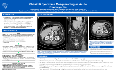
Labs revealed normal LFTs, lipase, lactate, and no pyuria. Ultrasound was negative for cholecystitis or cholelithiasis. CT of the abdomen and pelvis showed interposition of gaseous-distended distal ileum between the liver and right upper anterior abdominal wall, but without wall thickening or adjacent fat stranding (Figure 1).
Chilaiditi sign complicated by CS was recognized and given concerns for early SBO, General Surgery was consulted. After admission and NG decompression, she passed the Gastrografin challenge and had bowel movements, halting necessity for laparoscopic exploration. She tolerated diet advancement and was discharged.
Discussion: Chilaiditi sign is an uncommon finding with an incidence of between 0.25 and 0.28% on imaging. CS most commonly involves the colonic hepatic flexure and transverse colon. Ileum involvement is seldomly reported in the literature. While this patient presented with symptoms suggestive of cholecystitis, early recognition of SBO in the context of CS likely prevented surgical intervention and improved outcomes. Taken together, while Chilaiditi sign is typically benign, expeditious recognition of CS is crucial to prevent possible complications.

Disclosures:
Bibek Saha, MD, Nayantara Coelho-Prabhu, MBBS, Zachary S. Kelm, MD, PhD, Daniel Cabrera, MD. P2670 - Chilaiditi Syndrome Masquerading as Acute Cholecystitis, ACG 2023 Annual Scientific Meeting Abstracts. Vancouver, BC, Canada: American College of Gastroenterology.
Mayo Clinic, Rochester, MN
Introduction: Chilaiditi sign is an incidental radiological finding where the colon, or rarely, the small bowel is interposed between the diaphragm and liver. When present along with bowel obstruction or gastrointestinal symptoms, it is considered Chilaiditi Syndrome (CS). We present a rare case of CS causing small bowel obstruction (SBO) masquerading as acute cholecystitis and highlight the importance of early diagnosis, and management.
Case Description/Methods: A 73-year-old female with a history of GERD presented to the ED with a 3-day history of severe right upper quadrant (RUQ) abdominal pain with radiation to the right shoulder, and right back and flank. It worsened postprandially, and when lying flat or onto her right side, but was constant otherwise. She reported nausea and difficulty taking a deep breath due to the pain, but denied fever, emesis, constipation, hematochezia, and dysuria. She had no significant history of abdominal surgeries or alcohol abuse.
Her vital signs were overall normal. On physical exam, when laid flat, she developed severe pain. There was considerable tenderness in the RUQ and epigastrium, but Murphy’s sign was negative. The presentation early in the encounter was suggestive for biliary disease, particularly acute cholecystitis, and this was followed by appropriate workup.
Labs revealed normal LFTs, lipase, lactate, and no pyuria. Ultrasound was negative for cholecystitis or cholelithiasis. CT of the abdomen and pelvis showed interposition of gaseous-distended distal ileum between the liver and right upper anterior abdominal wall, but without wall thickening or adjacent fat stranding (Figure 1).
Chilaiditi sign complicated by CS was recognized and given concerns for early SBO, General Surgery was consulted. After admission and NG decompression, she passed the Gastrografin challenge and had bowel movements, halting necessity for laparoscopic exploration. She tolerated diet advancement and was discharged.
Discussion: Chilaiditi sign is an uncommon finding with an incidence of between 0.25 and 0.28% on imaging. CS most commonly involves the colonic hepatic flexure and transverse colon. Ileum involvement is seldomly reported in the literature. While this patient presented with symptoms suggestive of cholecystitis, early recognition of SBO in the context of CS likely prevented surgical intervention and improved outcomes. Taken together, while Chilaiditi sign is typically benign, expeditious recognition of CS is crucial to prevent possible complications.

Figure: Figure 1. Computed Tomography of the abdomen and pelvis showing interposition of gaseous-distended distal ileum (arrow) between the liver and the right upper anterior abdominal wall (Chilaiditi sign) but without wall thickening or adjacent fat stranding. A) Axial view. B) Coronal view.
Disclosures:
Bibek Saha indicated no relevant financial relationships.
Nayantara Coelho-Prabhu: Alexion Pharma – Advisor or Review Panel Member. Iterative Health – Advisor or Review Panel Member.
Zachary Kelm indicated no relevant financial relationships.
Daniel Cabrera indicated no relevant financial relationships.
Bibek Saha, MD, Nayantara Coelho-Prabhu, MBBS, Zachary S. Kelm, MD, PhD, Daniel Cabrera, MD. P2670 - Chilaiditi Syndrome Masquerading as Acute Cholecystitis, ACG 2023 Annual Scientific Meeting Abstracts. Vancouver, BC, Canada: American College of Gastroenterology.
