Monday Poster Session
Category: Small Intestine
P2689 - Behind the Pain: Unveiling an Uncommon Prothrombin Gene Mutation Leading to Devastating Bowel Ischemia
Monday, October 23, 2023
10:30 AM - 4:15 PM PT
Location: Exhibit Hall

Has Audio

Prateek S. Harne, MBBS, MD
University of Texas
Houston, TX
Presenting Author(s)
Prateek S. Harne, MBBS, MD1, Arturo Suplee Rivera, MD2, Shiv Yogesh. Govindji, MS2, Henry Herrera, MD3, Asif Zamir, MD, FACG4
1University of Texas, Houston, TX; 2University of Texas, Edinburg, TX; 3DHR Health, Edinburg, TX; 4DHR Health Gastroenterology, Edinburg, TX
Introduction: The rare prothrombin G20210A gene variant (PGV) is typically asymptomatic but can cause specific symptoms depending on the location and extent of thrombus. Extensive portal and superior mesenteric vein thrombosis resulting from this mutation is rarely documented. We present a case of severe bowel ischemia in a patient with the PGV, presenting initially as mild abdominal discomfort.
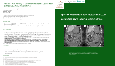
Case Description/Methods: A 34-year-old female with Down syndrome presented to her PCP with 10-day epigastric pain, diarrhea, rectal bleeding, and vomiting. CT scan showed jejunal thickening and inflammatory changes suggesting enteritis. Symptoms persisted, leading to hospital admission. Stool panel was negative. Repeat CT scan confirmed previous findings. Push enteroscopy and colonoscopy were unrevealing. She was readmitted with worsening symptoms, tachycardia (125) and leukocytosis (32,000), normal lactic acid. She received broad-spectrum antibiotics for presumed intra-abdominal infection. CT angiography revealed extensive mesenteric and portal vein thrombosis, enteritis, mesenteric edema, jejunal fluid distention, and moderate ascites. Exploratory laparotomy found large amounts of hemorrhagic ascites, grossly hemorrhagic necrosis of the mid-small bowel. Interestingly, hypercoagulable workup revealed PGV mutation without family history or risk factors. Recurrent mesenteric hemorrhage led to multiple surgical interventions (abdominal washout, ~200cm small bowel resection, wound-vac therapy, entero-enteral anastomosis). On day 30, discharged, symptom-free, and tolerating Apixaban during outpatient follow-up.
Discussion: Mesenteric ischemia and bowel necrosis secondary to extensive portal and superior mesenteric vein thrombosis caused by sporadic PGV mutation present scarcely reported. A family history or a triggering event such as pregnancy or surgery precedes the thrombotic event, however our case defied this pattern. CT abdomen with contrast may miss thrombi in inflammation, favoring CT angiography for diagnosis. This case underscores the importance of clinical awareness, timely evaluation, and intervention to prevent complications from mesenteric ischemia.

Disclosures:
Prateek S. Harne, MBBS, MD1, Arturo Suplee Rivera, MD2, Shiv Yogesh. Govindji, MS2, Henry Herrera, MD3, Asif Zamir, MD, FACG4. P2689 - Behind the Pain: Unveiling an Uncommon Prothrombin Gene Mutation Leading to Devastating Bowel Ischemia, ACG 2023 Annual Scientific Meeting Abstracts. Vancouver, BC, Canada: American College of Gastroenterology.
1University of Texas, Houston, TX; 2University of Texas, Edinburg, TX; 3DHR Health, Edinburg, TX; 4DHR Health Gastroenterology, Edinburg, TX
Introduction: The rare prothrombin G20210A gene variant (PGV) is typically asymptomatic but can cause specific symptoms depending on the location and extent of thrombus. Extensive portal and superior mesenteric vein thrombosis resulting from this mutation is rarely documented. We present a case of severe bowel ischemia in a patient with the PGV, presenting initially as mild abdominal discomfort.
Case Description/Methods: A 34-year-old female with Down syndrome presented to her PCP with 10-day epigastric pain, diarrhea, rectal bleeding, and vomiting. CT scan showed jejunal thickening and inflammatory changes suggesting enteritis. Symptoms persisted, leading to hospital admission. Stool panel was negative. Repeat CT scan confirmed previous findings. Push enteroscopy and colonoscopy were unrevealing. She was readmitted with worsening symptoms, tachycardia (125) and leukocytosis (32,000), normal lactic acid. She received broad-spectrum antibiotics for presumed intra-abdominal infection. CT angiography revealed extensive mesenteric and portal vein thrombosis, enteritis, mesenteric edema, jejunal fluid distention, and moderate ascites. Exploratory laparotomy found large amounts of hemorrhagic ascites, grossly hemorrhagic necrosis of the mid-small bowel. Interestingly, hypercoagulable workup revealed PGV mutation without family history or risk factors. Recurrent mesenteric hemorrhage led to multiple surgical interventions (abdominal washout, ~200cm small bowel resection, wound-vac therapy, entero-enteral anastomosis). On day 30, discharged, symptom-free, and tolerating Apixaban during outpatient follow-up.
Discussion: Mesenteric ischemia and bowel necrosis secondary to extensive portal and superior mesenteric vein thrombosis caused by sporadic PGV mutation present scarcely reported. A family history or a triggering event such as pregnancy or surgery precedes the thrombotic event, however our case defied this pattern. CT abdomen with contrast may miss thrombi in inflammation, favoring CT angiography for diagnosis. This case underscores the importance of clinical awareness, timely evaluation, and intervention to prevent complications from mesenteric ischemia.

Figure: Coronal section of CT Angiography (A and B) showing extensive mesenteric and portal vein thrombosis, enteritis, mesenteric edema, jejunal fluid distention, and moderate ascites.
Disclosures:
Prateek Harne indicated no relevant financial relationships.
Arturo Suplee Rivera indicated no relevant financial relationships.
Shiv Govindji indicated no relevant financial relationships.
Henry Herrera indicated no relevant financial relationships.
Asif Zamir indicated no relevant financial relationships.
Prateek S. Harne, MBBS, MD1, Arturo Suplee Rivera, MD2, Shiv Yogesh. Govindji, MS2, Henry Herrera, MD3, Asif Zamir, MD, FACG4. P2689 - Behind the Pain: Unveiling an Uncommon Prothrombin Gene Mutation Leading to Devastating Bowel Ischemia, ACG 2023 Annual Scientific Meeting Abstracts. Vancouver, BC, Canada: American College of Gastroenterology.
