Monday Poster Session
Category: Small Intestine
P2698 - The Constricted Voyage: A Case Report of a Retained Video Capsule from Strictures Caused by Focal Segmental Ischemia
Monday, October 23, 2023
10:30 AM - 4:15 PM PT
Location: Exhibit Hall

Has Audio

Abbas Bader
University of Missouri Kansas City
Kansas City, MO
Presenting Author(s)
Vinay Jahagirdar, MD1, Abbas Bader, 2, Adel Muhana, MD1, Kamani Lankachandra, MD1, Tariku Woldey, MD1, Rajiv Chhabra, MD3
1University of Missouri-Kansas City, Kansas City, MO; 2University of Missouri Kansas City, Kansas City, MO; 3University of Missouri-Kansas City School of Medicine, Saint Luke’s Hospital of Kansas City, Kansas City, MO
Introduction: Video capsule endoscopy (VCE) is widely used for evaluation of occult GI bleeding. Capsule retention, a rare but serious adverse event, is reported in < 1%. Small bowel blockage with a video capsule, in pre-existing stenosis, may result in dreaded obstruction. Stenosis may be due to context of underlying Crohn’s disease (CD), radiation therapy, bowel tumors or NSAID use. We report a case of capsule retention due to stricture from focal segmental ischemia of the small bowel, ultimately needing resection.
Case Description/Methods: A 49-year-old male with a history of Barrett's esophagus presented to GI clinic for management of refractory iron deficiency anemia (IDA). Recent bidirectional endoscopies were unrevealing. A VCE was done to further evaluate the IDA (PillCam SB3). The study was ineffective, as the capsule remained in the stomach for most of the duration and didn’t pass in the stool. Subsequent KUB showed the capsule within the RLQ. Patients denied any abdominal pain or obstructive symptoms.
3 weeks later he presented to the ED with worsening sharp RLQ pain for a day. Abdomen was tender with guarding. A CT abdomen revealed a possible pill camera, retained in the mid jejunum, along with diffuse small bowel thickening and dilation, and enlarged mesenteric lymph nodes. Due to capsule location, a double balloon anterograde enteroscopy was attempted for retrieval. However, it was unsuccessful due to strictures in the mid jejunum that could not be traversed. Patient eventually underwent laparoscopic small bowel resection to remove the capsule.
Pathology from resected small bowel was consistent with subacute/chronic ischemia with superimposed acute ischemia. Given the lack of other diagnostic findings for CD and the histopathological picture, the stricture was attributed to chronic focal segmental ischemia. On follow-up 11 months later, the patient was doing well.
Discussion: Manifestations of focal segmental ischemia of the small bowel, where short segments are affected, vary depending on the infarct severity. Causes of nonocclusive ischemic bowel disease include small vessel disease (connective tissue disorders, diabetes), hypoperfusion (hypercoagulability, shock, heart failure), or compromised blood flow from increased intraluminal pressure (constipation). Repeated ischemic injury may cause chronic enteritis which is difficult to differentiate from CD. Late stricture formation is also reported. Definitive diagnosis is by tissue biopsy, and surgical treatment by resection is often warranted.

Disclosures:
Vinay Jahagirdar, MD1, Abbas Bader, 2, Adel Muhana, MD1, Kamani Lankachandra, MD1, Tariku Woldey, MD1, Rajiv Chhabra, MD3. P2698 - The Constricted Voyage: A Case Report of a Retained Video Capsule from Strictures Caused by Focal Segmental Ischemia, ACG 2023 Annual Scientific Meeting Abstracts. Vancouver, BC, Canada: American College of Gastroenterology.
1University of Missouri-Kansas City, Kansas City, MO; 2University of Missouri Kansas City, Kansas City, MO; 3University of Missouri-Kansas City School of Medicine, Saint Luke’s Hospital of Kansas City, Kansas City, MO
Introduction: Video capsule endoscopy (VCE) is widely used for evaluation of occult GI bleeding. Capsule retention, a rare but serious adverse event, is reported in < 1%. Small bowel blockage with a video capsule, in pre-existing stenosis, may result in dreaded obstruction. Stenosis may be due to context of underlying Crohn’s disease (CD), radiation therapy, bowel tumors or NSAID use. We report a case of capsule retention due to stricture from focal segmental ischemia of the small bowel, ultimately needing resection.
Case Description/Methods: A 49-year-old male with a history of Barrett's esophagus presented to GI clinic for management of refractory iron deficiency anemia (IDA). Recent bidirectional endoscopies were unrevealing. A VCE was done to further evaluate the IDA (PillCam SB3). The study was ineffective, as the capsule remained in the stomach for most of the duration and didn’t pass in the stool. Subsequent KUB showed the capsule within the RLQ. Patients denied any abdominal pain or obstructive symptoms.
3 weeks later he presented to the ED with worsening sharp RLQ pain for a day. Abdomen was tender with guarding. A CT abdomen revealed a possible pill camera, retained in the mid jejunum, along with diffuse small bowel thickening and dilation, and enlarged mesenteric lymph nodes. Due to capsule location, a double balloon anterograde enteroscopy was attempted for retrieval. However, it was unsuccessful due to strictures in the mid jejunum that could not be traversed. Patient eventually underwent laparoscopic small bowel resection to remove the capsule.
Pathology from resected small bowel was consistent with subacute/chronic ischemia with superimposed acute ischemia. Given the lack of other diagnostic findings for CD and the histopathological picture, the stricture was attributed to chronic focal segmental ischemia. On follow-up 11 months later, the patient was doing well.
Discussion: Manifestations of focal segmental ischemia of the small bowel, where short segments are affected, vary depending on the infarct severity. Causes of nonocclusive ischemic bowel disease include small vessel disease (connective tissue disorders, diabetes), hypoperfusion (hypercoagulability, shock, heart failure), or compromised blood flow from increased intraluminal pressure (constipation). Repeated ischemic injury may cause chronic enteritis which is difficult to differentiate from CD. Late stricture formation is also reported. Definitive diagnosis is by tissue biopsy, and surgical treatment by resection is often warranted.

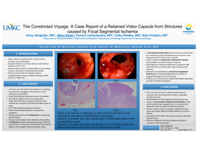
Figure: A,B: Stenosis in distal jejunum with a stricture that could not be traversed on antegrade double-balloon enteroscopy. C: Low power view (20X magnification) of the small bowel segment with mucosal inflammation. D:Low power view (40X magnification) of the small bowel mucosa with acute inflammation and overlying acute inflammatory exudate
Disclosures:
Vinay Jahagirdar indicated no relevant financial relationships.
Abbas Bader indicated no relevant financial relationships.
Adel Muhana indicated no relevant financial relationships.
Kamani Lankachandra indicated no relevant financial relationships.
Tariku Woldey indicated no relevant financial relationships.
Rajiv Chhabra indicated no relevant financial relationships.
Vinay Jahagirdar, MD1, Abbas Bader, 2, Adel Muhana, MD1, Kamani Lankachandra, MD1, Tariku Woldey, MD1, Rajiv Chhabra, MD3. P2698 - The Constricted Voyage: A Case Report of a Retained Video Capsule from Strictures Caused by Focal Segmental Ischemia, ACG 2023 Annual Scientific Meeting Abstracts. Vancouver, BC, Canada: American College of Gastroenterology.
