Monday Poster Session
Category: Small Intestine
P2707 - When a Polyp Isn't Really a Polyp: Duodenal Lymphangioma
Monday, October 23, 2023
10:30 AM - 4:15 PM PT
Location: Exhibit Hall

Has Audio

Jose F. Nunez-Morales, MD
VA Caribbean Healthcare System
San Juan, PR
Presenting Author(s)
Jose F.. Nunez-Morales, MD, Hector Nieves-Figueroa, MD, Giovanni Rivera-Colon, MD, Zeyn Mirza, MD, Andres Rabell-Bernal, MD
VA Caribbean Healthcare System, San Juan, Puerto Rico
Introduction: Lymphangiomas are rare, but benign malformations of the lymphatic system. They develop secondary to failure of individual lymphatic tissue to connect with the general lymphatic system. Usually, they present in the neck, head, and axilla. Lymphangiomas of the GI tract are rare, and ones at the duodenum are even more infrequent. They are mostly asymptomatic and usually diagnosed incidentally by EGD. Here we report a case of a duodenal lymphangioma which was found incidentally by EGD and confirmed by biopsy.
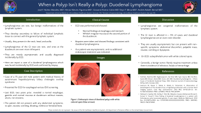
Case Description/Methods: Case of a 75-year-old male patient with medical history of suspected autoimmune hepatitis/primary biliary cholangitis overlap syndrome who presented for EGD for esophageal varices (EV) screening. Last EGD, two years prior, revealed a normal esophagus, gastritis, and normal mucosa at duodenum without masses, ulcers, nor erosions. The patient did not present with any abdominal symptoms, no pain, nausea, vomiting, bloating, melena, or hematochezia. EGD was performed and showed normal findings at esophagus and stomach but revealed a whitish irregular mucosa at the second portion of the duodenum. Biopsies were taken and showed findings consistent with duodenal lymphangioma.
Discussion: Lymphangiomas are congenital malformations of the lymphatic system. The GI tract is only affected in less than 5% of cases and duodenal lymphangiomas are an even rarer disorder. They are usually asymptomatic but can present with non-specific symptoms such as abdominal discomfort, palpable mass, nausea, vomiting or dyspepsia. On endoscopy, they can present as submucosal tumors with white colored spots. They are generally benign tumor that rarely requires treatment unless accompanied by infection, fistula, or hemorrhage. Our patient was asymptomatic, and no additional endoscopic treatment was indicated as would have been the case in a similar appearing small duodenal tubular adenoma.

Disclosures:
Jose F.. Nunez-Morales, MD, Hector Nieves-Figueroa, MD, Giovanni Rivera-Colon, MD, Zeyn Mirza, MD, Andres Rabell-Bernal, MD. P2707 - When a Polyp Isn't Really a Polyp: Duodenal Lymphangioma, ACG 2023 Annual Scientific Meeting Abstracts. Vancouver, BC, Canada: American College of Gastroenterology.
VA Caribbean Healthcare System, San Juan, Puerto Rico
Introduction: Lymphangiomas are rare, but benign malformations of the lymphatic system. They develop secondary to failure of individual lymphatic tissue to connect with the general lymphatic system. Usually, they present in the neck, head, and axilla. Lymphangiomas of the GI tract are rare, and ones at the duodenum are even more infrequent. They are mostly asymptomatic and usually diagnosed incidentally by EGD. Here we report a case of a duodenal lymphangioma which was found incidentally by EGD and confirmed by biopsy.
Case Description/Methods: Case of a 75-year-old male patient with medical history of suspected autoimmune hepatitis/primary biliary cholangitis overlap syndrome who presented for EGD for esophageal varices (EV) screening. Last EGD, two years prior, revealed a normal esophagus, gastritis, and normal mucosa at duodenum without masses, ulcers, nor erosions. The patient did not present with any abdominal symptoms, no pain, nausea, vomiting, bloating, melena, or hematochezia. EGD was performed and showed normal findings at esophagus and stomach but revealed a whitish irregular mucosa at the second portion of the duodenum. Biopsies were taken and showed findings consistent with duodenal lymphangioma.
Discussion: Lymphangiomas are congenital malformations of the lymphatic system. The GI tract is only affected in less than 5% of cases and duodenal lymphangiomas are an even rarer disorder. They are usually asymptomatic but can present with non-specific symptoms such as abdominal discomfort, palpable mass, nausea, vomiting or dyspepsia. On endoscopy, they can present as submucosal tumors with white colored spots. They are generally benign tumor that rarely requires treatment unless accompanied by infection, fistula, or hemorrhage. Our patient was asymptomatic, and no additional endoscopic treatment was indicated as would have been the case in a similar appearing small duodenal tubular adenoma.

Figure: EGD showing whitish irregular mucosa at the second portion of the duodenum.
Disclosures:
Jose Nunez-Morales indicated no relevant financial relationships.
Hector Nieves-Figueroa indicated no relevant financial relationships.
Giovanni Rivera-Colon indicated no relevant financial relationships.
Zeyn Mirza indicated no relevant financial relationships.
Andres Rabell-Bernal indicated no relevant financial relationships.
Jose F.. Nunez-Morales, MD, Hector Nieves-Figueroa, MD, Giovanni Rivera-Colon, MD, Zeyn Mirza, MD, Andres Rabell-Bernal, MD. P2707 - When a Polyp Isn't Really a Polyp: Duodenal Lymphangioma, ACG 2023 Annual Scientific Meeting Abstracts. Vancouver, BC, Canada: American College of Gastroenterology.
