Monday Poster Session
Category: Stomach
P2770 - Gas Everywhere! A Rare Case of Candida-Associated Emphysematous Gastritis
Monday, October 23, 2023
10:30 AM - 4:15 PM PT
Location: Exhibit Hall

Has Audio
- KM
Kalyana Y. Mondreti, MD
University of Texas Rio Grande Valley-DHR
McAllen, TX
Presenting Author(s)
Kalyana Y. Mondreti, MD1, Nghia Nguyen, DO1, Asif Zamir, MD, FACG2, Subrahmanyam Behara, MD3
1University of Texas Rio Grande Valley-DHR, Edinburg, TX; 2DHR Health Gastroenterology, Edinburg, TX; 3Doctors Hospital at Renaissance, Edinburg, TX
Introduction: Emphysematous Gastritis (EG) is an exceedingly rare and lethal condition characterized by gas in the gastric mucosal wall with diffuse inflammation. Only 59 cases have been reported and no management guidelines are currently available. Here we present a rare case of EG associated with Candida which is only the second case reported.
Case Description/Methods: A 41-year-old lady with a history of diabetes, and colectomy due to recurrent Clostridioides difficile presented with nausea, vomiting, and weakness. Upon arrival, her vitals were stable. Initial laboratories were remarkable for diabetic ketoacidosis (DKA) and urinary tract infection (UTI). CT scan of the abdomen showed mild distal esophageal distension and a retroperitoneal soft tissue mass. She was treated for DKA and UTI. However, 3 days later, the abdominal pain, nausea, and vomiting worsened despite the resolution of DKA. Labs showed WBC 14, Hemoglobin 9, and Lactate 2.7. Repeat CT of the abdomen showed new widespread ischemic small bowel with severe pneumatosis, presence of gas in the gastric mucosal wall along with superior mesenteric and portal veins. An emergent exploratory laparotomy showed a diffusely dilated small bowel with no ischemic changes and an indurated retroperitoneal mass. The mass was resected, and the abdomen was temporarily closed with a wound vac. Next day, the patient developed shock with bloody drainage from the NG tube. An emergent EGD showed a distended gastric body with extensive ulcerations and necrosis involving the stomach and duodenum. Biopsies from these lesions showed evidence of ischemic gastritis with Candida species. Micafungin was started and a second look laparotomy showed large areas of necrotic small bowel, which led to 21-cm small bowel resection and anastomosis. Afterward, the patient’s clinical condition improved significantly.
Discussion: EG is a rare entity characterized by the presence of gas in the gastric mucosal wall which can also cause portal venous gas as in our case. Mortality is around 60%, going up to 75% in surgical patients. Common organisms causing EG are E. coli, Pseudomonas, and Clostridioides but rarely Candida. CT abdomen is the imaging of choice, but an early EGD can help with a preliminary diagnosis. Supportive care and the prompt initiation of antifungal therapy are the mainstays of treatment. Surgery is indicated for patients with hemodynamic instability or perforation.

Disclosures:
Kalyana Y. Mondreti, MD1, Nghia Nguyen, DO1, Asif Zamir, MD, FACG2, Subrahmanyam Behara, MD3. P2770 - Gas Everywhere! A Rare Case of Candida-Associated Emphysematous Gastritis, ACG 2023 Annual Scientific Meeting Abstracts. Vancouver, BC, Canada: American College of Gastroenterology.
1University of Texas Rio Grande Valley-DHR, Edinburg, TX; 2DHR Health Gastroenterology, Edinburg, TX; 3Doctors Hospital at Renaissance, Edinburg, TX
Introduction: Emphysematous Gastritis (EG) is an exceedingly rare and lethal condition characterized by gas in the gastric mucosal wall with diffuse inflammation. Only 59 cases have been reported and no management guidelines are currently available. Here we present a rare case of EG associated with Candida which is only the second case reported.
Case Description/Methods: A 41-year-old lady with a history of diabetes, and colectomy due to recurrent Clostridioides difficile presented with nausea, vomiting, and weakness. Upon arrival, her vitals were stable. Initial laboratories were remarkable for diabetic ketoacidosis (DKA) and urinary tract infection (UTI). CT scan of the abdomen showed mild distal esophageal distension and a retroperitoneal soft tissue mass. She was treated for DKA and UTI. However, 3 days later, the abdominal pain, nausea, and vomiting worsened despite the resolution of DKA. Labs showed WBC 14, Hemoglobin 9, and Lactate 2.7. Repeat CT of the abdomen showed new widespread ischemic small bowel with severe pneumatosis, presence of gas in the gastric mucosal wall along with superior mesenteric and portal veins. An emergent exploratory laparotomy showed a diffusely dilated small bowel with no ischemic changes and an indurated retroperitoneal mass. The mass was resected, and the abdomen was temporarily closed with a wound vac. Next day, the patient developed shock with bloody drainage from the NG tube. An emergent EGD showed a distended gastric body with extensive ulcerations and necrosis involving the stomach and duodenum. Biopsies from these lesions showed evidence of ischemic gastritis with Candida species. Micafungin was started and a second look laparotomy showed large areas of necrotic small bowel, which led to 21-cm small bowel resection and anastomosis. Afterward, the patient’s clinical condition improved significantly.
Discussion: EG is a rare entity characterized by the presence of gas in the gastric mucosal wall which can also cause portal venous gas as in our case. Mortality is around 60%, going up to 75% in surgical patients. Common organisms causing EG are E. coli, Pseudomonas, and Clostridioides but rarely Candida. CT abdomen is the imaging of choice, but an early EGD can help with a preliminary diagnosis. Supportive care and the prompt initiation of antifungal therapy are the mainstays of treatment. Surgery is indicated for patients with hemodynamic instability or perforation.

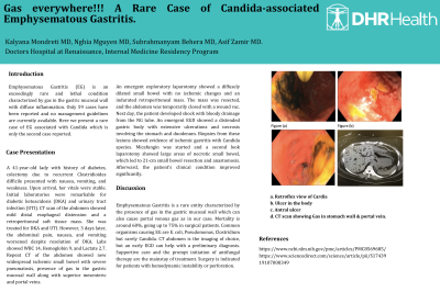
Figure: a. Retroflex view of Cardia
b. Ulcer in the body
c. Antral ulcer
d. CT scan showing Gas in stomach wall & portal vein.
b. Ulcer in the body
c. Antral ulcer
d. CT scan showing Gas in stomach wall & portal vein.
Disclosures:
Kalyana Mondreti indicated no relevant financial relationships.
Nghia Nguyen indicated no relevant financial relationships.
Asif Zamir indicated no relevant financial relationships.
Subrahmanyam Behara indicated no relevant financial relationships.
Kalyana Y. Mondreti, MD1, Nghia Nguyen, DO1, Asif Zamir, MD, FACG2, Subrahmanyam Behara, MD3. P2770 - Gas Everywhere! A Rare Case of Candida-Associated Emphysematous Gastritis, ACG 2023 Annual Scientific Meeting Abstracts. Vancouver, BC, Canada: American College of Gastroenterology.
