Monday Poster Session
Category: Stomach
P2782 - A Rare Case of Hemorrhagic Shock Secondary to Gastric Lipoma!
Monday, October 23, 2023
10:30 AM - 4:15 PM PT
Location: Exhibit Hall

Has Audio

Tahseen Azeem, DO
University Hospitals Parma Medical Center
Parma, OH
Presenting Author(s)
Tahseen Azeem, DO1, Sohaib Hassan, DO1, Adrian S. Lindsey, MD2, Fady G. Haddad, MD3
1University Hospitals Parma Medical Center, Parma, OH; 2CWRU/UH/VA, Cleveland, OH; 3University Hospitals, Parma, OH
Introduction: Lipomas are rare benign tumors formed of adipose tissue. When in the gastrointestinal tract (GI), lipomas are most commonly found in the colon and small bowel with only < 5% reported in the stomach. Despite that gastric lipomas are described, it is extremely uncommon for them to present with significant upper GI bleeding. Herein we report the case of an elderly patient presenting with hematemesis found to be secondary to a gastric lipoma.
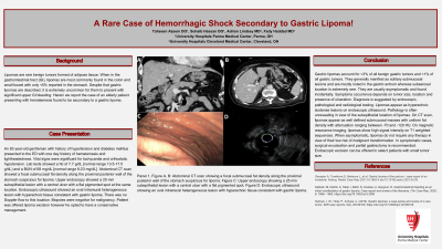
Case Description/Methods: An 83 year-old gentleman with history of hypertension and diabetes mellitus presented to the ED with 1 day history of hematemesis and lightheadedness. Vital signs were significant for tachycardia and orthostatic hypotension. Lab tests showed a hb of 7.7 g/dL [normal range 13.5-17.5 g/dL) and a BUN of 68 mg/dL [normal range 6-23 mg/dL]. Abdominal CT scan showed a focal submucosal fat density along the proximal posterior wall of the stomach suspicious for lipoma. Upper endoscopy showed a 25 mm subepithelial lesion with a central ulcer with a flat pigmented spot at the same location. Endoscopic ultrasound showed an oval intramural heterogeneous lesion with hyperechoic tissue consistent with gastric lipoma. There was no Doppler flow to this location. Biopsies were negative for malignancy. Patient was offered lipoma excision however he opted to have a conservative management.
Discussion: Gastric lipomas account for < 3% of all benign gastric tumors and < 1% of all gastric tumors. They generally manifest as solitary submucosal lesions and are mostly noted in the gastric antrum whereas subserosal location is extremely rare. They are usually asymptomatic and found incidentally. Symptoms occurrence depends on tumor size, location and presence of ulceration. Diagnosis is suggested by endoscopic, pathological and radiological testing. Lipomas appear as hyperechoic isodense lesions on endoscopic ultrasound. Pathology is often unrevealing in view of the subepithelial location of lipomas. On CT scan, lipomas appear as well defined submucosal masses with uniform fat density with attenuation ranging between -70 and -120 HU. On magnetic resonance imaging, lipomas show high signal intensity on T1 weighted sequences. When asymptomatic, lipomas do not require any therapy in view of their low risk of malignant transformation. In symptomatic cases, surgical enucleation and partial gastrectomy is recommended. Endoscopic excision can be offered in select patients with small tumor size.

Disclosures:
Tahseen Azeem, DO1, Sohaib Hassan, DO1, Adrian S. Lindsey, MD2, Fady G. Haddad, MD3. P2782 - A Rare Case of Hemorrhagic Shock Secondary to Gastric Lipoma!, ACG 2023 Annual Scientific Meeting Abstracts. Vancouver, BC, Canada: American College of Gastroenterology.
1University Hospitals Parma Medical Center, Parma, OH; 2CWRU/UH/VA, Cleveland, OH; 3University Hospitals, Parma, OH
Introduction: Lipomas are rare benign tumors formed of adipose tissue. When in the gastrointestinal tract (GI), lipomas are most commonly found in the colon and small bowel with only < 5% reported in the stomach. Despite that gastric lipomas are described, it is extremely uncommon for them to present with significant upper GI bleeding. Herein we report the case of an elderly patient presenting with hematemesis found to be secondary to a gastric lipoma.
Case Description/Methods: An 83 year-old gentleman with history of hypertension and diabetes mellitus presented to the ED with 1 day history of hematemesis and lightheadedness. Vital signs were significant for tachycardia and orthostatic hypotension. Lab tests showed a hb of 7.7 g/dL [normal range 13.5-17.5 g/dL) and a BUN of 68 mg/dL [normal range 6-23 mg/dL]. Abdominal CT scan showed a focal submucosal fat density along the proximal posterior wall of the stomach suspicious for lipoma. Upper endoscopy showed a 25 mm subepithelial lesion with a central ulcer with a flat pigmented spot at the same location. Endoscopic ultrasound showed an oval intramural heterogeneous lesion with hyperechoic tissue consistent with gastric lipoma. There was no Doppler flow to this location. Biopsies were negative for malignancy. Patient was offered lipoma excision however he opted to have a conservative management.
Discussion: Gastric lipomas account for < 3% of all benign gastric tumors and < 1% of all gastric tumors. They generally manifest as solitary submucosal lesions and are mostly noted in the gastric antrum whereas subserosal location is extremely rare. They are usually asymptomatic and found incidentally. Symptoms occurrence depends on tumor size, location and presence of ulceration. Diagnosis is suggested by endoscopic, pathological and radiological testing. Lipomas appear as hyperechoic isodense lesions on endoscopic ultrasound. Pathology is often unrevealing in view of the subepithelial location of lipomas. On CT scan, lipomas appear as well defined submucosal masses with uniform fat density with attenuation ranging between -70 and -120 HU. On magnetic resonance imaging, lipomas show high signal intensity on T1 weighted sequences. When asymptomatic, lipomas do not require any therapy in view of their low risk of malignant transformation. In symptomatic cases, surgical enucleation and partial gastrectomy is recommended. Endoscopic excision can be offered in select patients with small tumor size.

Figure: Panel 1, Figure A, B: Abdominal CT scan showing a focal submucosal fat density along the proximal posterior wall of the stomach suspicious for lipoma; Figure C: Upper endoscopy showing a 25 mm subepithelial lesion with a central ulcer with a flat pigmented spot; Figure D: Endoscopic ultrasound showing an oval intramural heterogeneous lesion with hyperechoic tissue consistent with gastric lipoma.
Disclosures:
Tahseen Azeem indicated no relevant financial relationships.
Sohaib Hassan indicated no relevant financial relationships.
Adrian Lindsey indicated no relevant financial relationships.
Fady G. Haddad indicated no relevant financial relationships.
Tahseen Azeem, DO1, Sohaib Hassan, DO1, Adrian S. Lindsey, MD2, Fady G. Haddad, MD3. P2782 - A Rare Case of Hemorrhagic Shock Secondary to Gastric Lipoma!, ACG 2023 Annual Scientific Meeting Abstracts. Vancouver, BC, Canada: American College of Gastroenterology.
