Monday Poster Session
Category: Stomach
P2784 - Malignant Gastrointestinal Stromal Tumor Presenting With Spontaneous Intraperitoneal Hemorrhage in a Patient on Dual Antiplatelet Therapy
Monday, October 23, 2023
10:30 AM - 4:15 PM PT
Location: Exhibit Hall

Has Audio

Sinay Ceballos, MD
HCA Aventura Hospital & Medical Center
Miami, FL
Presenting Author(s)
Sinay Ceballos, MD1, Daniel Campbell, DO2, Arash Zarrin, DO2, Luis G.. Santiago Zayas, MD2, Shany M.. Quevedo, MD3, Ramasamy Nathan, MD2, Franklin Kasmin, MD2
1HCA Aventura Hospital & Medical Center, Miami, FL; 2HCA Florida Aventura Hospital, Aventura, FL; 3HCA East Florida Division, Aventura, FL
Introduction: Gastrointestinal stromal tumors (GIST) are rare mesenchymal neoplasms of the gastrointestinal tract. They represent approximately 1-2% of all gastrointestinal cancer and the most common mesenchymal neoplasm. GISTs can arise from any portion of the gastrointestinal tract, with up to 60% originating from the stomach. They are predominantly seen in elderly patients. Here we present an interesting case about a patient with abdominal pain secondary to a large highly vascularized mass, presumably a GIST.
Case Description/Methods: A 73-year-old male with history of CAD on DAPT, H. pylori (+) chronic gastritis and a Forrest class IIB duodenal ulcer status post clipping (2016) presented with a 4-day history of severe sharp pain in the left hemiabdomen, nausea and watery non-bloody bowel movements. Hemodynamically stable on admission. Initial labs showed leukocytosis and a NCNC anemia with Hgb 12.8. A CT angiography of the abdomen and pelvis showed a large gastrointestinal stromal tumor (GIST) arising from the gastric wall (Figure 1) with an intraperitoneal hemorrhage. No luminal gastrointestinal bleed was found . A celiac angiogram showed innumerable small branches from the splenic artery supplying the mass, with no active extravasation. Embolization was not feasible due to extensive blood supply. EGD showed a normal gastric and duodenal mucosa and Endoscopic ultrasound (EUS) demonstrated a large doppler negative perigastric mass with no pathologic lymph nodes. Pathology from specimens collected through FNA reported a low-grade GIST. Patient improved clinically and was subsequently discharged, currently undergoing medical/surgical oncologic treatment.
Discussion: The clinical presentation for GIST is variable. Most tumors originate from the stomach and are diagnosed after an overt or occult gastrointestinal bleed. Our patient developed severe abdominal pain and was found to have a large gastric wall tumor with abundant vascular supply causing self-limited intraperitoneal bleed. In most cases due to the risk of intraabdominal bleed, tumor embolization is attempted, especially in patients on anticoagulation or antiplatelet therapy, which unfortunately was unsuccessful in our patient. EUS allow us to better assess tumor extension and to obtain specimens for pathology confirmation. A thorough exam, supportive imaging and access to medical procedures facilitate early diagnosis of patients with GISTs, directed to improve their outcome.

Disclosures:
Sinay Ceballos, MD1, Daniel Campbell, DO2, Arash Zarrin, DO2, Luis G.. Santiago Zayas, MD2, Shany M.. Quevedo, MD3, Ramasamy Nathan, MD2, Franklin Kasmin, MD2. P2784 - Malignant Gastrointestinal Stromal Tumor Presenting With Spontaneous Intraperitoneal Hemorrhage in a Patient on Dual Antiplatelet Therapy, ACG 2023 Annual Scientific Meeting Abstracts. Vancouver, BC, Canada: American College of Gastroenterology.
1HCA Aventura Hospital & Medical Center, Miami, FL; 2HCA Florida Aventura Hospital, Aventura, FL; 3HCA East Florida Division, Aventura, FL
Introduction: Gastrointestinal stromal tumors (GIST) are rare mesenchymal neoplasms of the gastrointestinal tract. They represent approximately 1-2% of all gastrointestinal cancer and the most common mesenchymal neoplasm. GISTs can arise from any portion of the gastrointestinal tract, with up to 60% originating from the stomach. They are predominantly seen in elderly patients. Here we present an interesting case about a patient with abdominal pain secondary to a large highly vascularized mass, presumably a GIST.
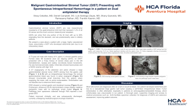
Case Description/Methods: A 73-year-old male with history of CAD on DAPT, H. pylori (+) chronic gastritis and a Forrest class IIB duodenal ulcer status post clipping (2016) presented with a 4-day history of severe sharp pain in the left hemiabdomen, nausea and watery non-bloody bowel movements. Hemodynamically stable on admission. Initial labs showed leukocytosis and a NCNC anemia with Hgb 12.8. A CT angiography of the abdomen and pelvis showed a large gastrointestinal stromal tumor (GIST) arising from the gastric wall (Figure 1) with an intraperitoneal hemorrhage. No luminal gastrointestinal bleed was found . A celiac angiogram showed innumerable small branches from the splenic artery supplying the mass, with no active extravasation. Embolization was not feasible due to extensive blood supply. EGD showed a normal gastric and duodenal mucosa and Endoscopic ultrasound (EUS) demonstrated a large doppler negative perigastric mass with no pathologic lymph nodes. Pathology from specimens collected through FNA reported a low-grade GIST. Patient improved clinically and was subsequently discharged, currently undergoing medical/surgical oncologic treatment.
Discussion: The clinical presentation for GIST is variable. Most tumors originate from the stomach and are diagnosed after an overt or occult gastrointestinal bleed. Our patient developed severe abdominal pain and was found to have a large gastric wall tumor with abundant vascular supply causing self-limited intraperitoneal bleed. In most cases due to the risk of intraabdominal bleed, tumor embolization is attempted, especially in patients on anticoagulation or antiplatelet therapy, which unfortunately was unsuccessful in our patient. EUS allow us to better assess tumor extension and to obtain specimens for pathology confirmation. A thorough exam, supportive imaging and access to medical procedures facilitate early diagnosis of patients with GISTs, directed to improve their outcome.

Figure: Figure 1:
CT angiography of the abdomen and pelvis, coronal view, showing a large mass possibly a GIST arising from the gastric wall measuring 11x14 cm. Noted significant interface with the splenic hilum.
CT angiography of the abdomen and pelvis, coronal view, showing a large mass possibly a GIST arising from the gastric wall measuring 11x14 cm. Noted significant interface with the splenic hilum.
Disclosures:
Sinay Ceballos indicated no relevant financial relationships.
Daniel Campbell indicated no relevant financial relationships.
Arash Zarrin indicated no relevant financial relationships.
Luis Santiago Zayas indicated no relevant financial relationships.
Shany Quevedo indicated no relevant financial relationships.
Ramasamy Nathan indicated no relevant financial relationships.
Franklin Kasmin indicated no relevant financial relationships.
Sinay Ceballos, MD1, Daniel Campbell, DO2, Arash Zarrin, DO2, Luis G.. Santiago Zayas, MD2, Shany M.. Quevedo, MD3, Ramasamy Nathan, MD2, Franklin Kasmin, MD2. P2784 - Malignant Gastrointestinal Stromal Tumor Presenting With Spontaneous Intraperitoneal Hemorrhage in a Patient on Dual Antiplatelet Therapy, ACG 2023 Annual Scientific Meeting Abstracts. Vancouver, BC, Canada: American College of Gastroenterology.
