Monday Poster Session
Category: Stomach
P2785 - Metastatic Melanoma Involving the Gastrointestinal Tract
Monday, October 23, 2023
10:30 AM - 4:15 PM PT
Location: Exhibit Hall

Has Audio

Neil Shah, MS, MD
Trinity Health Ann Arbor
Ann Arbor, MI
Presenting Author(s)
Neil Shah, MS, MD1, Kais Antonios, MD1, Priyata Dutta, MD2
1Trinity Health Ann Arbor, Ann Arbor, MI; 2Trinity Health Ann Arbor, Ypsilanti, MI
Introduction: Malignant melanoma is one of the leading causes of mortality related to skin cancer. The GI tract is commonly involved; however, the majority of GI metastasis is discovered post-mortem. While the jejunum, ileum, rectum and colon are the most common sites of metastasis, gastric metastasis is rare and underreported.
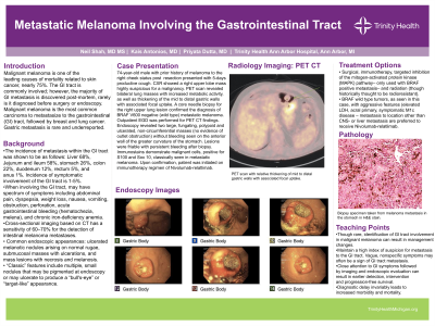
Case Description/Methods: 74-year-old male with a distant personal medical history of melanoma to the right cheek status post surgical resection presented to the clinic due to a 5-day period of a productive cough. A CXR showed a right upper lobe mass highly suspicious for a malignancy. PET scan revealed bilateral lung masses with increased metabolic activity, compatible with malignancy as well as relative thickening of the mid to distal gastric walls with associated focal uptake (Figure 1). A core needle biopsy for the right upper lung lesion confirmed the diagnosis of BRAF V600 negative (wild type) metastatic melanoma. Outpatient EGD was performed for the above mentioned PET CT findings. Endoscopy revealed two large, fungating, polypoid and ulcerated, non-circumferential masses (no evidence of outlet obstruction) with no bleeding seen on the anterior wall of the greater curvature of the stomach. Lesions were extremely friable with persistent bleeding after biopsy (Figure 2). Immunostains demonstrate malignant cells, positive for S100 and Sox 10, classically seen in metastatic melanoma. Upon confirmation of metastatic melanoma to the stomach and lungs, the patient was initiated on immunotherapy regimen of Nivolumab and Relatlimab-rmbw.
Discussion: Although metastatic melanoma to the GI tract is common, gastric metastasis is unusual, and usually not discovered until late in the disease course. In the case described, our patient did not present with any GI manifestations, rather abnormal findings were seen on advanced cross sectional imaging. Diagnostic delay invariably leads to increased morbidity and mortality. Therefore, close attention to GI symptoms followed by imaging and endoscopic evaluation can result in earlier intervention and progression-free survival.

Disclosures:
Neil Shah, MS, MD1, Kais Antonios, MD1, Priyata Dutta, MD2. P2785 - Metastatic Melanoma Involving the Gastrointestinal Tract, ACG 2023 Annual Scientific Meeting Abstracts. Vancouver, BC, Canada: American College of Gastroenterology.
1Trinity Health Ann Arbor, Ann Arbor, MI; 2Trinity Health Ann Arbor, Ypsilanti, MI
Introduction: Malignant melanoma is one of the leading causes of mortality related to skin cancer. The GI tract is commonly involved; however, the majority of GI metastasis is discovered post-mortem. While the jejunum, ileum, rectum and colon are the most common sites of metastasis, gastric metastasis is rare and underreported.
Case Description/Methods: 74-year-old male with a distant personal medical history of melanoma to the right cheek status post surgical resection presented to the clinic due to a 5-day period of a productive cough. A CXR showed a right upper lobe mass highly suspicious for a malignancy. PET scan revealed bilateral lung masses with increased metabolic activity, compatible with malignancy as well as relative thickening of the mid to distal gastric walls with associated focal uptake (Figure 1). A core needle biopsy for the right upper lung lesion confirmed the diagnosis of BRAF V600 negative (wild type) metastatic melanoma. Outpatient EGD was performed for the above mentioned PET CT findings. Endoscopy revealed two large, fungating, polypoid and ulcerated, non-circumferential masses (no evidence of outlet obstruction) with no bleeding seen on the anterior wall of the greater curvature of the stomach. Lesions were extremely friable with persistent bleeding after biopsy (Figure 2). Immunostains demonstrate malignant cells, positive for S100 and Sox 10, classically seen in metastatic melanoma. Upon confirmation of metastatic melanoma to the stomach and lungs, the patient was initiated on immunotherapy regimen of Nivolumab and Relatlimab-rmbw.
Discussion: Although metastatic melanoma to the GI tract is common, gastric metastasis is unusual, and usually not discovered until late in the disease course. In the case described, our patient did not present with any GI manifestations, rather abnormal findings were seen on advanced cross sectional imaging. Diagnostic delay invariably leads to increased morbidity and mortality. Therefore, close attention to GI symptoms followed by imaging and endoscopic evaluation can result in earlier intervention and progression-free survival.

Figure: Left: PET scan with relative thickening of the mid to distal gastric walls with associated focal uptake
Right: EGD findings of two ulcerated polypoid masses in the anterior wall of the stomach measuring 4cm (top) and 1.5cm (bottom)
Right: EGD findings of two ulcerated polypoid masses in the anterior wall of the stomach measuring 4cm (top) and 1.5cm (bottom)
Disclosures:
Neil Shah indicated no relevant financial relationships.
Kais Antonios indicated no relevant financial relationships.
Priyata Dutta indicated no relevant financial relationships.
Neil Shah, MS, MD1, Kais Antonios, MD1, Priyata Dutta, MD2. P2785 - Metastatic Melanoma Involving the Gastrointestinal Tract, ACG 2023 Annual Scientific Meeting Abstracts. Vancouver, BC, Canada: American College of Gastroenterology.

