Monday Poster Session
Category: Stomach
P2803 - When Your World Gets Turned Upside Down
Monday, October 23, 2023
10:30 AM - 4:15 PM PT
Location: Exhibit Hall

Has Audio

Patricia Rivera, MD
VA Caribbean Healthcare System
San Juan, PR
Presenting Author(s)
Patricia Rivera, MD, Orlando Rodriguez-Amador, MD, Carolina Diaz-loza, MD, Jaime Martinez-Souss, MD
VA Caribbean Healthcare System, San Juan, Puerto Rico
Introduction: Gastric volvulus is a rare, but life-threatening condition that results from the abnormal rotation of the stomach around one of its axes beyond 180 degrees. If not treated promptly or misdiagnosed it may result in gastric strangulation and necrosis. Among the different types of gastric volvulus, organoaxial is the most common, seen in up to 60% of the cases, where the rotation of the stomach occurs along its long axis. Despite being the most common precipitating factor of this condition, only 4% of hiatal hernias are complicated by gastric volvulus. Here we present the case of an acute organoaxial gastric volvulus due to a longstanding massive hiatal hernia.
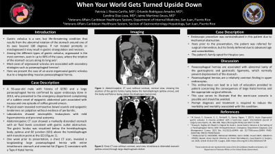
Case Description/Methods: A 96-year-old male with history of GERD and a large hiatal hernia, diagnosed in 2013 via upper endoscopy, who presented to the emergency department complaining of a sudden onset of epigastric abdominal pain associated with nausea and one episode of coffee ground emesis. Physical exam revealed normoactive bowel sounds and epigastric tenderness on palpation without evidence of peritonitis. Laboratories showed neutrophilic leukocytosis with mild hypernatremia and pre-renal azotemia. Abdominopelvic CT scan showed a markedly distended stomach with air fluid levels consistent with gastric outlet obstruction. The gastric fundus was visualized below the hemidiaphragm; body, pylorus and GE junction (GEJ) above the hemidiaphragm with transition point at the GEJ (Figure 1). One week prior to presentation, a chest CT scan, revealed a longstanding large hiatal hernia with entire intrathoracic stomach and omental fat (Figure 2) consistent with a Type 4 hiatal hernia. Endoscopic procedure was contraindicated in this patient in the setting of mechanical obstruction. Years prior to the presentation, the patient was referred for surgical intervention but his family deferred due to advanced age and comorbidities. The patient’s family opted for Hospice care.
Discussion: There are several factors that may contribute to the development of a hiatal hernia, including advanced age and intra-abdominal pressure. Hiatal hernias are a relatively common finding in upper endoscopies. This ordinariness can lead to a lack of education provided to patient concerning the consequences of large hiatal hernias and the appropriate surgical referrals. This case serves to illustrate that the worst case scenario is possible and should be avoided. Prompt diagnosis and treatment is required to reduce the morbidity and mortality associated with this condition.

Disclosures:
Patricia Rivera, MD, Orlando Rodriguez-Amador, MD, Carolina Diaz-loza, MD, Jaime Martinez-Souss, MD. P2803 - When Your World Gets Turned Upside Down, ACG 2023 Annual Scientific Meeting Abstracts. Vancouver, BC, Canada: American College of Gastroenterology.
VA Caribbean Healthcare System, San Juan, Puerto Rico
Introduction: Gastric volvulus is a rare, but life-threatening condition that results from the abnormal rotation of the stomach around one of its axes beyond 180 degrees. If not treated promptly or misdiagnosed it may result in gastric strangulation and necrosis. Among the different types of gastric volvulus, organoaxial is the most common, seen in up to 60% of the cases, where the rotation of the stomach occurs along its long axis. Despite being the most common precipitating factor of this condition, only 4% of hiatal hernias are complicated by gastric volvulus. Here we present the case of an acute organoaxial gastric volvulus due to a longstanding massive hiatal hernia.
Case Description/Methods: A 96-year-old male with history of GERD and a large hiatal hernia, diagnosed in 2013 via upper endoscopy, who presented to the emergency department complaining of a sudden onset of epigastric abdominal pain associated with nausea and one episode of coffee ground emesis. Physical exam revealed normoactive bowel sounds and epigastric tenderness on palpation without evidence of peritonitis. Laboratories showed neutrophilic leukocytosis with mild hypernatremia and pre-renal azotemia. Abdominopelvic CT scan showed a markedly distended stomach with air fluid levels consistent with gastric outlet obstruction. The gastric fundus was visualized below the hemidiaphragm; body, pylorus and GE junction (GEJ) above the hemidiaphragm with transition point at the GEJ (Figure 1). One week prior to presentation, a chest CT scan, revealed a longstanding large hiatal hernia with entire intrathoracic stomach and omental fat (Figure 2) consistent with a Type 4 hiatal hernia. Endoscopic procedure was contraindicated in this patient in the setting of mechanical obstruction. Years prior to the presentation, the patient was referred for surgical intervention but his family deferred due to advanced age and comorbidities. The patient’s family opted for Hospice care.
Discussion: There are several factors that may contribute to the development of a hiatal hernia, including advanced age and intra-abdominal pressure. Hiatal hernias are a relatively common finding in upper endoscopies. This ordinariness can lead to a lack of education provided to patient concerning the consequences of large hiatal hernias and the appropriate surgical referrals. This case serves to illustrate that the worst case scenario is possible and should be avoided. Prompt diagnosis and treatment is required to reduce the morbidity and mortality associated with this condition.

Figure: Figure 1: Abdominopelvic CT scan without contrast, coronal view showing the position of the gastric fundus laying below the hemidiaphragm (A), and the body and Pylorus laying above the diaphragm (B).
Figure 2: Chest CT scan without contrast, axial view. Intrathoracic distended stomach (A) through large hiatal hernia defect.
Figure 2: Chest CT scan without contrast, axial view. Intrathoracic distended stomach (A) through large hiatal hernia defect.
Disclosures:
Patricia Rivera indicated no relevant financial relationships.
Orlando Rodriguez-Amador indicated no relevant financial relationships.
Carolina Diaz-loza indicated no relevant financial relationships.
Jaime Martinez-Souss indicated no relevant financial relationships.
Patricia Rivera, MD, Orlando Rodriguez-Amador, MD, Carolina Diaz-loza, MD, Jaime Martinez-Souss, MD. P2803 - When Your World Gets Turned Upside Down, ACG 2023 Annual Scientific Meeting Abstracts. Vancouver, BC, Canada: American College of Gastroenterology.
