Monday Poster Session
Category: Stomach
P2812 - Gastric Lipoma: A Rare Cause of Melena
Monday, October 23, 2023
10:30 AM - 4:15 PM PT
Location: Exhibit Hall

Has Audio

Alexandria Markley, MD
Icahn School of Medicine at Mount Sinai
New York, NY
Presenting Author(s)
Alexandria Markley, MD1, Rohit Nathani, MD2, Erica Park, MD3, Armand Cacciarelli, MD4
1Icahn School of Medicine at Mount Sinai, New York, NY; 2Mount Sinai Morningside and West Hospital, New York, NY; 3Mount Sinai Morningside and West, New York, NY; 4Mount Sinai, New York, NY
Introduction: Gastric lipomas (GL) are rare benign tumors, accounting for less than 1% of all gastric tumors arising from adipose tissue within the stomach. They are often asymptomatic and slow growing but can cause symptoms and complications such as gastric outlet obstruction and gastrointestinal (GI) bleeding. We present a case of a GL incidentally discovered during an esophagogastroduodenoscopy (EGD) for melena.
Case Description/Methods: A 66-year-old male with a medical history of hypertension, benign prostate hyperplasia, and obesity presented with dark stools. Physical examination was unremarkable and initial laboratory studies revealed iron deficiency anemia with a Hb of 9.8 mg/dl. The differential diagnosis was broad for causes of upper GI bleeding and the patient underwent EGD. EGD revealed a medium-sized, non-circumferential, ulcerated mass in the gastric antrum without active bleeding. Histology confirmed a gastric hyperplastic polyp with no malignancy. Further evaluation with endoscopic ultrasound (EUS) identified an intramural, hyperechoic lesion originating from the submucosa. Pathology showed fibroadipose tissue with reactive features consistent with an ulcerated lipoma. Due to its large size, the patient was referred for surgical evaluation. Computed Tomography (CT) scan showed a 9.1 x 4.5cm fat density lesion. The patient underwent diagnostic laparoscopy, robotic subtotal gastrectomy with removal of perigastric lymph nodes, longitudinal gastrotomy, and excision of intragastric mass. The mass was confirmed to be a well differentiated lipoma on excisional biopsy. The patient had complete resolution of symptoms.
Discussion: GL are often asymptomatic; however, in some instances, as described above, GL can cause symptoms such as abdominal pain, bloating, nausea, vomiting, or GI bleeding if it is associated with ulceration or necrosis.
Initial evaluation with EGD allows direct visualization of the lesion. Imaging techniques such as EUS, CT, or magnetic resonance imaging can further provide valuable information about its size, location, and appearance.
Small asymptomatic GL can be managed conservatively with regular surveillance EGDs. GL under 4 cm can typically be removed endoscopically. However, larger lipomas or symptomatic cases, particularly those with concerns of ulceration, bleeding, or diagnostic uncertainty, may require surgical removal. Recurrence is rare post-removal, and long-term follow-up is generally unnecessary.

Disclosures:
Alexandria Markley, MD1, Rohit Nathani, MD2, Erica Park, MD3, Armand Cacciarelli, MD4. P2812 - Gastric Lipoma: A Rare Cause of Melena, ACG 2023 Annual Scientific Meeting Abstracts. Vancouver, BC, Canada: American College of Gastroenterology.
1Icahn School of Medicine at Mount Sinai, New York, NY; 2Mount Sinai Morningside and West Hospital, New York, NY; 3Mount Sinai Morningside and West, New York, NY; 4Mount Sinai, New York, NY
Introduction: Gastric lipomas (GL) are rare benign tumors, accounting for less than 1% of all gastric tumors arising from adipose tissue within the stomach. They are often asymptomatic and slow growing but can cause symptoms and complications such as gastric outlet obstruction and gastrointestinal (GI) bleeding. We present a case of a GL incidentally discovered during an esophagogastroduodenoscopy (EGD) for melena.
Case Description/Methods: A 66-year-old male with a medical history of hypertension, benign prostate hyperplasia, and obesity presented with dark stools. Physical examination was unremarkable and initial laboratory studies revealed iron deficiency anemia with a Hb of 9.8 mg/dl. The differential diagnosis was broad for causes of upper GI bleeding and the patient underwent EGD. EGD revealed a medium-sized, non-circumferential, ulcerated mass in the gastric antrum without active bleeding. Histology confirmed a gastric hyperplastic polyp with no malignancy. Further evaluation with endoscopic ultrasound (EUS) identified an intramural, hyperechoic lesion originating from the submucosa. Pathology showed fibroadipose tissue with reactive features consistent with an ulcerated lipoma. Due to its large size, the patient was referred for surgical evaluation. Computed Tomography (CT) scan showed a 9.1 x 4.5cm fat density lesion. The patient underwent diagnostic laparoscopy, robotic subtotal gastrectomy with removal of perigastric lymph nodes, longitudinal gastrotomy, and excision of intragastric mass. The mass was confirmed to be a well differentiated lipoma on excisional biopsy. The patient had complete resolution of symptoms.
Discussion: GL are often asymptomatic; however, in some instances, as described above, GL can cause symptoms such as abdominal pain, bloating, nausea, vomiting, or GI bleeding if it is associated with ulceration or necrosis.
Initial evaluation with EGD allows direct visualization of the lesion. Imaging techniques such as EUS, CT, or magnetic resonance imaging can further provide valuable information about its size, location, and appearance.
Small asymptomatic GL can be managed conservatively with regular surveillance EGDs. GL under 4 cm can typically be removed endoscopically. However, larger lipomas or symptomatic cases, particularly those with concerns of ulceration, bleeding, or diagnostic uncertainty, may require surgical removal. Recurrence is rare post-removal, and long-term follow-up is generally unnecessary.

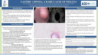
Figure: A. Upper Gastrointestinal Endoscopy image showing large subepithelial gastric antrum mass with ulceration. B. Endoscopic Ultrasound image demonstrating an intramural lesion in the antrum of the stomach.
Disclosures:
Alexandria Markley indicated no relevant financial relationships.
Rohit Nathani indicated no relevant financial relationships.
Erica Park indicated no relevant financial relationships.
Armand Cacciarelli indicated no relevant financial relationships.
Alexandria Markley, MD1, Rohit Nathani, MD2, Erica Park, MD3, Armand Cacciarelli, MD4. P2812 - Gastric Lipoma: A Rare Cause of Melena, ACG 2023 Annual Scientific Meeting Abstracts. Vancouver, BC, Canada: American College of Gastroenterology.

