Monday Poster Session
Category: Stomach
P2827 - A Rare Case of Gastric Leiomyosarcoma With Peculiar Endoscopic Appearance
Monday, October 23, 2023
10:30 AM - 4:15 PM PT
Location: Exhibit Hall

Has Audio

Nevil Kadakia, MD
One Brooklyn Health-Interfaith Medical Center
Ozone Park, NY
Presenting Author(s)
Nevil Kadakia, MD1, Pratik Munishwar, MD2, Uma D. Gupta, MD3, Derrick Cheung, MD4, Benjamin Silver, MD4, Elliot Bigajer, MD5
1One Brooklyn Health-Interfaith Medical Center, Queens, NY; 2Krakenhaus Winsen, Winsen, Niedersachsen, Germany; 3One Brooklyn Health-Interfaith Medical Center, Brooklyn, NY; 4One Brooklyn Health, Brooklyn, NY; 5One Brooklyn Health-Brookdale University Hospital Medical Center, Brooklyn, NY
Introduction: Primary leiomyosarcomas of the stomach are extremely rare. Historically, several cases of leiomyosarcomas of the stomach were misclassified as gastrointestinal stromal tumors (GISTs) prior to the advent of KIT immunohistochemistry (KIT era). Due to the low incidence of this tumor, clinicians still have not formed a therapeutic consensus. In this report, we present an intriguing case of a gastric leiomyosarcoma with a distinctive endoscopic appearance, thereby contributing this rare entity to the existing literature.
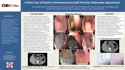
Case Description/Methods: A 49-year-old male, presented to the emergency department with a three-week history of nausea, vomiting, poor appetite, abdominal pain and weight loss. An initial computed tomography scan of the abdomen and pelvis with intravenous contrast revealed a large mid to distal gastric mass with marked wall thickening and central necrosis/air-fluid level, measuring 7 x 5 cm in size. The differential diagnoses included a necrotic gastric lymphoma vs adenocarcinoma. Additionally, numerous peri-gastric lymph nodes and hepatic lesions were observed, indicating potential metastases. An esophagogastroduodenoscopy demonstrated a large, cratered ulcer with heaped-up edges in the distal stomach, invading the duodenal bulb. Biopsies from the ulcer edge was inconclusive. An interventional radiology (IR) guided liver biopsy was performed which diagnosed a leiomyosarcoma, with positive immunohistochemical stains for Smooth Muscle Actin (SMA) and Desmin, while KI-67 revealed a high proliferative index. Conversely, stains for cd34, cd117, dog1, bcl2, and s100 were negative. These findings excluded the possibility of a gastrointestinal stromal tumor (GIST) and supported the diagnosis of a smooth muscle neoplasm.
Discussion: Leiomyosarcomas are aggressive tumors characterized by a positive immunohistochemical stain for actin and desmin. They also have a negative stain for CD117, CD34, and DOG1, thereby differentiating a leiomyosarcoma from a GIST. Accurate identification and pathological differentiation between the two are crucial, as GISTs demonstrate an excellent response to targeted treatment with tyrosine kinase inhibitors (TKIs). In our case, a previously healthy patient with no medical history was diagnosed with a rare leiomyosarcoma. The treatment plan involved tumor resection and adjuvant chemotherapy as determined by the surgery and oncology team.

Disclosures:
Nevil Kadakia, MD1, Pratik Munishwar, MD2, Uma D. Gupta, MD3, Derrick Cheung, MD4, Benjamin Silver, MD4, Elliot Bigajer, MD5. P2827 - A Rare Case of Gastric Leiomyosarcoma With Peculiar Endoscopic Appearance, ACG 2023 Annual Scientific Meeting Abstracts. Vancouver, BC, Canada: American College of Gastroenterology.
1One Brooklyn Health-Interfaith Medical Center, Queens, NY; 2Krakenhaus Winsen, Winsen, Niedersachsen, Germany; 3One Brooklyn Health-Interfaith Medical Center, Brooklyn, NY; 4One Brooklyn Health, Brooklyn, NY; 5One Brooklyn Health-Brookdale University Hospital Medical Center, Brooklyn, NY
Introduction: Primary leiomyosarcomas of the stomach are extremely rare. Historically, several cases of leiomyosarcomas of the stomach were misclassified as gastrointestinal stromal tumors (GISTs) prior to the advent of KIT immunohistochemistry (KIT era). Due to the low incidence of this tumor, clinicians still have not formed a therapeutic consensus. In this report, we present an intriguing case of a gastric leiomyosarcoma with a distinctive endoscopic appearance, thereby contributing this rare entity to the existing literature.
Case Description/Methods: A 49-year-old male, presented to the emergency department with a three-week history of nausea, vomiting, poor appetite, abdominal pain and weight loss. An initial computed tomography scan of the abdomen and pelvis with intravenous contrast revealed a large mid to distal gastric mass with marked wall thickening and central necrosis/air-fluid level, measuring 7 x 5 cm in size. The differential diagnoses included a necrotic gastric lymphoma vs adenocarcinoma. Additionally, numerous peri-gastric lymph nodes and hepatic lesions were observed, indicating potential metastases. An esophagogastroduodenoscopy demonstrated a large, cratered ulcer with heaped-up edges in the distal stomach, invading the duodenal bulb. Biopsies from the ulcer edge was inconclusive. An interventional radiology (IR) guided liver biopsy was performed which diagnosed a leiomyosarcoma, with positive immunohistochemical stains for Smooth Muscle Actin (SMA) and Desmin, while KI-67 revealed a high proliferative index. Conversely, stains for cd34, cd117, dog1, bcl2, and s100 were negative. These findings excluded the possibility of a gastrointestinal stromal tumor (GIST) and supported the diagnosis of a smooth muscle neoplasm.
Discussion: Leiomyosarcomas are aggressive tumors characterized by a positive immunohistochemical stain for actin and desmin. They also have a negative stain for CD117, CD34, and DOG1, thereby differentiating a leiomyosarcoma from a GIST. Accurate identification and pathological differentiation between the two are crucial, as GISTs demonstrate an excellent response to targeted treatment with tyrosine kinase inhibitors (TKIs). In our case, a previously healthy patient with no medical history was diagnosed with a rare leiomyosarcoma. The treatment plan involved tumor resection and adjuvant chemotherapy as determined by the surgery and oncology team.

Figure: Advances in immunohistochemistry led to the decrease of the incidence of gastric leiomyosarcomas to 1% of all malignant gastric tumors. Endoscopy unable to retroflexed of the stomach and absence of a discernible duodenal bulb
Disclosures:
Nevil Kadakia indicated no relevant financial relationships.
Pratik Munishwar indicated no relevant financial relationships.
Uma Gupta indicated no relevant financial relationships.
Derrick Cheung indicated no relevant financial relationships.
Benjamin Silver indicated no relevant financial relationships.
Elliot Bigajer indicated no relevant financial relationships.
Nevil Kadakia, MD1, Pratik Munishwar, MD2, Uma D. Gupta, MD3, Derrick Cheung, MD4, Benjamin Silver, MD4, Elliot Bigajer, MD5. P2827 - A Rare Case of Gastric Leiomyosarcoma With Peculiar Endoscopic Appearance, ACG 2023 Annual Scientific Meeting Abstracts. Vancouver, BC, Canada: American College of Gastroenterology.
