Monday Poster Session
Category: Stomach
P2833 - Gastric Linitis Plastica - An Unusual Presentation of Metastatic Breast Cancer
Monday, October 23, 2023
10:30 AM - 4:15 PM PT
Location: Exhibit Hall

Has Audio
- AV
Adam Vinall, MD
University of Texas Health Science Center at San Antonio
San Antonio, TX
Presenting Author(s)
Adam Vinall, MD1, James Alvarez, MD2, Chandraprakash Umapathy, MD, MS1
1University of Texas Health Science Center at San Antonio, San Antonio, TX; 2University of Texas Health Science Center-San Antonio, San Antonio, TX
Introduction: In women, breast cancer is the most common malignancy and the second most common cause of cancer death. It is typically discovered by screening mammography or a palpable breast mass on physical exam. Here we present an unusual case of metastatic breast cancer diagnosed by esophagogastroduodenoscopy (EGD) during the work up of gastroesophageal reflux disease (GERD) with alarm features.
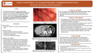
Case Description/Methods: A 59-year-old female with no past medical history presented for evaluation of heartburn and nausea. She had been tried on famotidine and ondansetron without relief for a presumed diagnosis of GERD. Review of systems was positive for intermittent hematochezia, constipation, a left-sided neck mass, and forty pounds of weight loss over the preceding three months. Physical exam was notable for a firm, mobile 4 cm mass of the left neck. Fecal immunochemical test was positive and H. pylori stool antigen was negative. A screening mammogram from 6 years prior was negative. An EGD was performed which showed gastric deformity concerning for linitis plastica (Figure 1c). A colonoscopy was unremarkable. Computed tomography (CT) of abdomen/pelvis showed gastric wall thickening (Figure 1b) and retroperitoneal, pelvic sidewall, and inguinal lymphadenopathy. CT chest showed left breast nodularity and follow up mammography showed multifocal disease in the left breast (Figure 1a). Imaging of the neck mass was suspicious for pathologic adenopathy. Biopsies showed poorly differentiated carcinoma consistent with lobular breast carcinoma. The carcinoma was strongly estrogen receptor, progesterone receptor, and Her2 positive. She was started on docetaxel, trastuzumab and pertuzumab with a good response.
Discussion: The most common sites for metastatic breast cancer are the bone, liver, and lungs. This case illustrates an unusual metastatic spread involving the stomach. The constellation of weight loss, heartburn, and palpable adenopathy in the left neck are classically concerning for gastric adenocarcinoma and prompt evaluation with endoscopy is vital for early diagnosis and management. Linitis plastica is also an endoscopic sign of diffuse and infiltrative gastric cancer where submucosal invasion leads to a characteristic “leathery” appearance. A similar radiographic and endoscopic appearance can be seen in metastatic cancer to the stomach. This case prompts us to have an additional diagnostic consideration when performing an evaluation of alarm features with endoscopy.

Disclosures:
Adam Vinall, MD1, James Alvarez, MD2, Chandraprakash Umapathy, MD, MS1. P2833 - Gastric Linitis Plastica - An Unusual Presentation of Metastatic Breast Cancer, ACG 2023 Annual Scientific Meeting Abstracts. Vancouver, BC, Canada: American College of Gastroenterology.
1University of Texas Health Science Center at San Antonio, San Antonio, TX; 2University of Texas Health Science Center-San Antonio, San Antonio, TX
Introduction: In women, breast cancer is the most common malignancy and the second most common cause of cancer death. It is typically discovered by screening mammography or a palpable breast mass on physical exam. Here we present an unusual case of metastatic breast cancer diagnosed by esophagogastroduodenoscopy (EGD) during the work up of gastroesophageal reflux disease (GERD) with alarm features.
Case Description/Methods: A 59-year-old female with no past medical history presented for evaluation of heartburn and nausea. She had been tried on famotidine and ondansetron without relief for a presumed diagnosis of GERD. Review of systems was positive for intermittent hematochezia, constipation, a left-sided neck mass, and forty pounds of weight loss over the preceding three months. Physical exam was notable for a firm, mobile 4 cm mass of the left neck. Fecal immunochemical test was positive and H. pylori stool antigen was negative. A screening mammogram from 6 years prior was negative. An EGD was performed which showed gastric deformity concerning for linitis plastica (Figure 1c). A colonoscopy was unremarkable. Computed tomography (CT) of abdomen/pelvis showed gastric wall thickening (Figure 1b) and retroperitoneal, pelvic sidewall, and inguinal lymphadenopathy. CT chest showed left breast nodularity and follow up mammography showed multifocal disease in the left breast (Figure 1a). Imaging of the neck mass was suspicious for pathologic adenopathy. Biopsies showed poorly differentiated carcinoma consistent with lobular breast carcinoma. The carcinoma was strongly estrogen receptor, progesterone receptor, and Her2 positive. She was started on docetaxel, trastuzumab and pertuzumab with a good response.
Discussion: The most common sites for metastatic breast cancer are the bone, liver, and lungs. This case illustrates an unusual metastatic spread involving the stomach. The constellation of weight loss, heartburn, and palpable adenopathy in the left neck are classically concerning for gastric adenocarcinoma and prompt evaluation with endoscopy is vital for early diagnosis and management. Linitis plastica is also an endoscopic sign of diffuse and infiltrative gastric cancer where submucosal invasion leads to a characteristic “leathery” appearance. A similar radiographic and endoscopic appearance can be seen in metastatic cancer to the stomach. This case prompts us to have an additional diagnostic consideration when performing an evaluation of alarm features with endoscopy.

Figure: (a) Left breast with diffuse calcifications and architectural distortions concerning for malignancy. (b) Diffuse gastric wall thickening with hyperenhancement was present throughout the entire stomach. (c) Deformity and poor distensibility in the gastric body, concerning for linitis plastica.
Disclosures:
Adam Vinall indicated no relevant financial relationships.
James Alvarez indicated no relevant financial relationships.
Chandraprakash Umapathy indicated no relevant financial relationships.
Adam Vinall, MD1, James Alvarez, MD2, Chandraprakash Umapathy, MD, MS1. P2833 - Gastric Linitis Plastica - An Unusual Presentation of Metastatic Breast Cancer, ACG 2023 Annual Scientific Meeting Abstracts. Vancouver, BC, Canada: American College of Gastroenterology.
