Monday Poster Session
Category: Stomach
P2837 - Plasmacytoma: A Rare Type of Gastric Submucosal Lesion
Monday, October 23, 2023
10:30 AM - 4:15 PM PT
Location: Exhibit Hall

Has Audio
- AL
Andy Lin, MD
University of California Irvine
Orange, California
Presenting Author(s)
Andy Lin, MD1, David L. Cheung, MD2, Amirali Tavangar, MD1, Samuel Ji, MD1, Thinh Pham, 1, Khaldoon Khirfan, MD1, Jason Samarasena, MD1
1University of California Irvine, Orange, CA; 2UC Irvine, Orange, CA
Introduction: Multiple Myeloma (MM) is a plasma cell neoplasm typically localized in the bone marrow and characterized by monoclonal protein production. At time of diagnosis, about 7 percent of patients will also have an extramedullary plasmacytoma (EP) which can be seen on Positron Emission Tomography or computed tomography, and 6 percent of patients will go on to develop an EP later in the disease course. We present a case of gastric plasmacytoma 2 years after MM diagnosis.
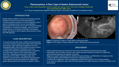
Case Description/Methods: A 76-year-old male with history of MM and plasmacytomas in the liver and spine presents with melena and acute blood loss anemia with a hemoglobin drop from 8.5 g/dL. Upper endoscopy revealed an over 5 cm subepithelial gastric lesion with superficial erosions (Figure 1). Endoscopic ultrasound confirmed an over 5 cm homogenous hypoechoic subepithelial lesion arising from the submucosa. (Figure 2) Fine needle biopsy was performed. Pathology showed plasma cells diffusely expressing CD138, MUM-1 with Kapp light chain restriction. Overall findings consistent with plasmacytoma. 15 Gy radiation therapy was performed to stabilize the gastric plasmacytoma with resolution of patient’s melena.
Discussion: Gastric plasmacytomas are the second most common EP following the head and neck region. Presentation can be variable, with prior case reports reporting nonspecific symptoms of weight loss, gastrointestinal bleeding and epigastric discomfort. Diagnosis of gastric plasmacytomas is definitive through pathology, as differentials on imaging can also include lymphoma, gastrointestinal stromal tumor, adenocarcinoma etc. If isolated EP is diagnosed, further investigations for MM should ensue as 15% of cases progress. In our case, EP appeared 2 years after the initial diagnosis of multiple myeloma, for which our patient is still being treated.

Disclosures:
Andy Lin, MD1, David L. Cheung, MD2, Amirali Tavangar, MD1, Samuel Ji, MD1, Thinh Pham, 1, Khaldoon Khirfan, MD1, Jason Samarasena, MD1. P2837 - Plasmacytoma: A Rare Type of Gastric Submucosal Lesion, ACG 2023 Annual Scientific Meeting Abstracts. Vancouver, BC, Canada: American College of Gastroenterology.
1University of California Irvine, Orange, CA; 2UC Irvine, Orange, CA
Introduction: Multiple Myeloma (MM) is a plasma cell neoplasm typically localized in the bone marrow and characterized by monoclonal protein production. At time of diagnosis, about 7 percent of patients will also have an extramedullary plasmacytoma (EP) which can be seen on Positron Emission Tomography or computed tomography, and 6 percent of patients will go on to develop an EP later in the disease course. We present a case of gastric plasmacytoma 2 years after MM diagnosis.
Case Description/Methods: A 76-year-old male with history of MM and plasmacytomas in the liver and spine presents with melena and acute blood loss anemia with a hemoglobin drop from 8.5 g/dL. Upper endoscopy revealed an over 5 cm subepithelial gastric lesion with superficial erosions (Figure 1). Endoscopic ultrasound confirmed an over 5 cm homogenous hypoechoic subepithelial lesion arising from the submucosa. (Figure 2) Fine needle biopsy was performed. Pathology showed plasma cells diffusely expressing CD138, MUM-1 with Kapp light chain restriction. Overall findings consistent with plasmacytoma. 15 Gy radiation therapy was performed to stabilize the gastric plasmacytoma with resolution of patient’s melena.
Discussion: Gastric plasmacytomas are the second most common EP following the head and neck region. Presentation can be variable, with prior case reports reporting nonspecific symptoms of weight loss, gastrointestinal bleeding and epigastric discomfort. Diagnosis of gastric plasmacytomas is definitive through pathology, as differentials on imaging can also include lymphoma, gastrointestinal stromal tumor, adenocarcinoma etc. If isolated EP is diagnosed, further investigations for MM should ensue as 15% of cases progress. In our case, EP appeared 2 years after the initial diagnosis of multiple myeloma, for which our patient is still being treated.

Figure: 1) Endoscopic image of a large gastric submucosal lesion
2) EUS image of a large hypoechoic lesion infiltrating the submucosal layer
2) EUS image of a large hypoechoic lesion infiltrating the submucosal layer
Disclosures:
Andy Lin indicated no relevant financial relationships.
David Cheung indicated no relevant financial relationships.
Amirali Tavangar indicated no relevant financial relationships.
Samuel Ji indicated no relevant financial relationships.
Thinh Pham indicated no relevant financial relationships.
Khaldoon Khirfan indicated no relevant financial relationships.
Jason Samarasena: Applied Medical – Advisor or Review Panel Member. Boston Scientific – Consultant. Conmed – Consultant. Cook – Educational Grant. Neptune Medical – Consultant. Olympus – Consultant. Steris – Consultant.
Andy Lin, MD1, David L. Cheung, MD2, Amirali Tavangar, MD1, Samuel Ji, MD1, Thinh Pham, 1, Khaldoon Khirfan, MD1, Jason Samarasena, MD1. P2837 - Plasmacytoma: A Rare Type of Gastric Submucosal Lesion, ACG 2023 Annual Scientific Meeting Abstracts. Vancouver, BC, Canada: American College of Gastroenterology.
