Monday Poster Session
Category: Liver
P2534 - Infectious Mimicry: A Diagnostic Conundrum in the Setting of Liver Lesions
Monday, October 23, 2023
10:30 AM - 4:15 PM PT
Location: Exhibit Hall

Has Audio

Jonathan Romanes, DO
Palmetto General Hospital
Pembroke Pines, FL
Presenting Author(s)
Jonathan Romanes, DO1, Adam Z. Koller, DO, MS2, Isabella Suarez Alcala, MD3, Roselys Ibanez Cabrera, MD4, Anita Bhagavathula, DO5
1Palmetto General Hospital, Pembroke Pines, FL; 2Palmetto General Hospital, Coral Gables, FL; 3Palmetto General Hospital, Hialeah, FL; 4Palmetto General hospital, Hialeah, FL; 5LCH South Miami, Doral, FL
Introduction: Differentiating between malignancy and infectious processes can be a diagnostic challenge, especially when clinical and radiologic findings overlap. Regarding liver lesions, the possibility of malignancy is often given more consideration. However, infectious processes can imitate malignant conditions, leading to diagnostic challenges. We present the case of a young male with weight loss, fever, and night sweats. Imaging revealed multiple liver lesions suspicious for metastatic disease, resulting in an extensive work-up for malignancy. After a thorough investigation, the true underlying etiology was unveiled.
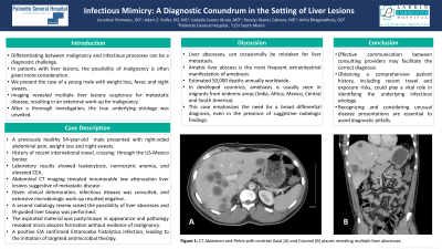
Case Description/Methods: A previously healthy male presented with right-sided abdominal pain, weight loss and night sweats. He also endorsed a history of recent international travel, crossing multiple countries while going through the US-Mexico border. Laboratory investigations showed leukocytosis, normocytic anemia, and elevated CEA. Abdominal CT imaging revealed innumerable low attenuation liver lesions suggestive of metastatic disease. However, given clinical deterioration, evaluation by infectious disease specialists was sought. Extensive laboratory investigations, including Gram stain, acid-fast bacilli, fungal culture, and serological assays, failed to identify a specific pathogen. A second radiology review raised the possibility of liver abscesses and IR-guided liver biopsy was performed. The aspirated material was pasty-brown in appearance and pathology revealed microabscess formation without evidence of malignancy. However, a positive EIA confirmed Entamoeba histolytica infection, leading to the initiation of targeted antimicrobial therapy. Given atypical presentation of liver abscesses, patient was scheduled to return for outpatient repeat CT scan imaging within 5 weeks to evaluate response to treatment.
Discussion: This case highlights several crucial points. First, the atypical presentation of an infectious process mimicking malignancy emphasizes the need for a broad differential diagnosis, even in the presence of suggestive radiologic findings. Second, effective communication between consulting providers, including radiologists and infectious disease specialists, facilitated the correct diagnosis. Third, obtaining a comprehensive patient history, including recent travel and exposure risks, played a vital role in identifying the underlying infectious etiology. Fourth, recognizing and considering unusual disease presentations are essential to avoid diagnostic pitfalls.

Disclosures:
Jonathan Romanes, DO1, Adam Z. Koller, DO, MS2, Isabella Suarez Alcala, MD3, Roselys Ibanez Cabrera, MD4, Anita Bhagavathula, DO5. P2534 - Infectious Mimicry: A Diagnostic Conundrum in the Setting of Liver Lesions, ACG 2023 Annual Scientific Meeting Abstracts. Vancouver, BC, Canada: American College of Gastroenterology.
1Palmetto General Hospital, Pembroke Pines, FL; 2Palmetto General Hospital, Coral Gables, FL; 3Palmetto General Hospital, Hialeah, FL; 4Palmetto General hospital, Hialeah, FL; 5LCH South Miami, Doral, FL
Introduction: Differentiating between malignancy and infectious processes can be a diagnostic challenge, especially when clinical and radiologic findings overlap. Regarding liver lesions, the possibility of malignancy is often given more consideration. However, infectious processes can imitate malignant conditions, leading to diagnostic challenges. We present the case of a young male with weight loss, fever, and night sweats. Imaging revealed multiple liver lesions suspicious for metastatic disease, resulting in an extensive work-up for malignancy. After a thorough investigation, the true underlying etiology was unveiled.
Case Description/Methods: A previously healthy male presented with right-sided abdominal pain, weight loss and night sweats. He also endorsed a history of recent international travel, crossing multiple countries while going through the US-Mexico border. Laboratory investigations showed leukocytosis, normocytic anemia, and elevated CEA. Abdominal CT imaging revealed innumerable low attenuation liver lesions suggestive of metastatic disease. However, given clinical deterioration, evaluation by infectious disease specialists was sought. Extensive laboratory investigations, including Gram stain, acid-fast bacilli, fungal culture, and serological assays, failed to identify a specific pathogen. A second radiology review raised the possibility of liver abscesses and IR-guided liver biopsy was performed. The aspirated material was pasty-brown in appearance and pathology revealed microabscess formation without evidence of malignancy. However, a positive EIA confirmed Entamoeba histolytica infection, leading to the initiation of targeted antimicrobial therapy. Given atypical presentation of liver abscesses, patient was scheduled to return for outpatient repeat CT scan imaging within 5 weeks to evaluate response to treatment.
Discussion: This case highlights several crucial points. First, the atypical presentation of an infectious process mimicking malignancy emphasizes the need for a broad differential diagnosis, even in the presence of suggestive radiologic findings. Second, effective communication between consulting providers, including radiologists and infectious disease specialists, facilitated the correct diagnosis. Third, obtaining a comprehensive patient history, including recent travel and exposure risks, played a vital role in identifying the underlying infectious etiology. Fourth, recognizing and considering unusual disease presentations are essential to avoid diagnostic pitfalls.

Figure: CT Abdomen and Pelvis with contrast showing innumerable liver abscesses
Disclosures:
Jonathan Romanes indicated no relevant financial relationships.
Adam Koller indicated no relevant financial relationships.
Isabella Suarez Alcala indicated no relevant financial relationships.
Roselys Ibanez Cabrera indicated no relevant financial relationships.
Anita Bhagavathula indicated no relevant financial relationships.
Jonathan Romanes, DO1, Adam Z. Koller, DO, MS2, Isabella Suarez Alcala, MD3, Roselys Ibanez Cabrera, MD4, Anita Bhagavathula, DO5. P2534 - Infectious Mimicry: A Diagnostic Conundrum in the Setting of Liver Lesions, ACG 2023 Annual Scientific Meeting Abstracts. Vancouver, BC, Canada: American College of Gastroenterology.
