Monday Poster Session
Category: Interventional Endoscopy
P2311 - Role of Endoscopic Ultrasound Fine Needle Biopsy Confirmation in a Large Retroperitoneal Mass
Monday, October 23, 2023
10:30 AM - 4:15 PM PT
Location: Exhibit Hall

Has Audio

Yash P. Ashara, MBBS
Mayo Clinic
Rochester, MN
Presenting Author(s)
Yash P. Ashara, MBBS1, Ruchir Paladiya, MBBS2, Allyson Pagan, MD3, Ami Bhalodia, MBBS4, Bhavtosh Dedania, MD5
1Mayo Clinic, Rochester, MN; 2UConn Health, Hartford, CT; 3HCA West Florida Brandon, Brandon, FL; 4Duke University, Durham, NC; 5HCA Florida Brandon Hospital, Brandon, FL
Introduction: Sarcomas are a rare group of malignant tumors derived from mesenchymal tissues, constituting less than 1 % of all adult malignancies. Retroperitoneal sarcoma is uncommon, comprising about 13% of all sarcomas. Here, we present a unique case of retroperitoneal well-differentiated liposarcoma diagnosed with endoscopic ultrasound (EUS) guided fine needle biopsy (FNB) and how it helped change the management in our patient.
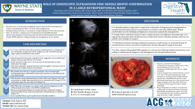
Case Description/Methods: A 77-year-old Thai male with a previous smoking history complained of loss of appetite, progressive 20 lb. weight loss over 6 months and left upper quadrant abdominal discomfort. CT of the abdomen revealed a large 19.5 x 13.2 cm mass in the LUQ with no clear distinction for origin. Labs showed Hb of 8.6 g/dL with MCV 74 fL suggestive of iron deficiency anemia with normal CEA, CA 19-9 & AFP. CTA of the abdomen showed extensive arterial supply to the mass, but no definitive site origin could be ascertained. The patient was seen by a surgical oncologist, who recommended undergoing biopsy confirmation before any resection, as the differentials were quite broad. EUS showed a large lobulated hyperechoic solid mass (Figure A-B) with no invasion or origin from pancreas, liver, or stomach wall. A large 23 X 22 mm lymph node was also observed (Figure C). EUS-FNB with a 22-gauge core needle confirmed the diagnosis of well-differentiated liposarcoma with MDM2 amplification. Based on pathology findings, the patient directly underwent surgical resection without any need for preoperative chemotherapy or radiation.
Discussion: Given the uncommon occurrence of liposarcoma and the complexities associated with its diagnosis and management, it is imperative to adopt a multidisciplinary approach for evaluation. A biopsy confirmation of the histological diagnosis is necessary as other differentials like a large GIST would warrant a neoadjuvant chemotherapy or a high-grade myxoid/round cell liposarcoma would need chemo-radiation therapy whereas our patient went straight to surgery. Traditional methods of image-guided percutaneous needle biopsy or incisional biopsy of retroperitoneal masses may pose complications during subsequent surgical resection. In this context, EUS-guided FNB emerges as a crucial tool, offering minimal invasiveness and aiding in confirming the diagnosis of retroperitoneal masses. This approach facilitates the identification of appropriate treatment strategies based on pathology findings.

Disclosures:
Yash P. Ashara, MBBS1, Ruchir Paladiya, MBBS2, Allyson Pagan, MD3, Ami Bhalodia, MBBS4, Bhavtosh Dedania, MD5. P2311 - Role of Endoscopic Ultrasound Fine Needle Biopsy Confirmation in a Large Retroperitoneal Mass, ACG 2023 Annual Scientific Meeting Abstracts. Vancouver, BC, Canada: American College of Gastroenterology.
1Mayo Clinic, Rochester, MN; 2UConn Health, Hartford, CT; 3HCA West Florida Brandon, Brandon, FL; 4Duke University, Durham, NC; 5HCA Florida Brandon Hospital, Brandon, FL
Introduction: Sarcomas are a rare group of malignant tumors derived from mesenchymal tissues, constituting less than 1 % of all adult malignancies. Retroperitoneal sarcoma is uncommon, comprising about 13% of all sarcomas. Here, we present a unique case of retroperitoneal well-differentiated liposarcoma diagnosed with endoscopic ultrasound (EUS) guided fine needle biopsy (FNB) and how it helped change the management in our patient.
Case Description/Methods: A 77-year-old Thai male with a previous smoking history complained of loss of appetite, progressive 20 lb. weight loss over 6 months and left upper quadrant abdominal discomfort. CT of the abdomen revealed a large 19.5 x 13.2 cm mass in the LUQ with no clear distinction for origin. Labs showed Hb of 8.6 g/dL with MCV 74 fL suggestive of iron deficiency anemia with normal CEA, CA 19-9 & AFP. CTA of the abdomen showed extensive arterial supply to the mass, but no definitive site origin could be ascertained. The patient was seen by a surgical oncologist, who recommended undergoing biopsy confirmation before any resection, as the differentials were quite broad. EUS showed a large lobulated hyperechoic solid mass (Figure A-B) with no invasion or origin from pancreas, liver, or stomach wall. A large 23 X 22 mm lymph node was also observed (Figure C). EUS-FNB with a 22-gauge core needle confirmed the diagnosis of well-differentiated liposarcoma with MDM2 amplification. Based on pathology findings, the patient directly underwent surgical resection without any need for preoperative chemotherapy or radiation.
Discussion: Given the uncommon occurrence of liposarcoma and the complexities associated with its diagnosis and management, it is imperative to adopt a multidisciplinary approach for evaluation. A biopsy confirmation of the histological diagnosis is necessary as other differentials like a large GIST would warrant a neoadjuvant chemotherapy or a high-grade myxoid/round cell liposarcoma would need chemo-radiation therapy whereas our patient went straight to surgery. Traditional methods of image-guided percutaneous needle biopsy or incisional biopsy of retroperitoneal masses may pose complications during subsequent surgical resection. In this context, EUS-guided FNB emerges as a crucial tool, offering minimal invasiveness and aiding in confirming the diagnosis of retroperitoneal masses. This approach facilitates the identification of appropriate treatment strategies based on pathology findings.

Figure: A: Large hyperechoic mass;
B: Fine Needle Biopsy of mass;
C: 23 x 21 mm lymph node
B: Fine Needle Biopsy of mass;
C: 23 x 21 mm lymph node
Disclosures:
Yash Ashara indicated no relevant financial relationships.
Ruchir Paladiya indicated no relevant financial relationships.
Allyson Pagan indicated no relevant financial relationships.
Ami Bhalodia indicated no relevant financial relationships.
Bhavtosh Dedania indicated no relevant financial relationships.
Yash P. Ashara, MBBS1, Ruchir Paladiya, MBBS2, Allyson Pagan, MD3, Ami Bhalodia, MBBS4, Bhavtosh Dedania, MD5. P2311 - Role of Endoscopic Ultrasound Fine Needle Biopsy Confirmation in a Large Retroperitoneal Mass, ACG 2023 Annual Scientific Meeting Abstracts. Vancouver, BC, Canada: American College of Gastroenterology.
