Monday Poster Session
Category: Esophagus
P1901 - Dysphagia Megalatriensis: A Cardiac Etiology Masquerading as Gastroesophageal Dysphagia
Monday, October 23, 2023
10:30 AM - 4:15 PM PT
Location: Exhibit Hall

Has Audio

Clive J. Miranda, DO, MS
University at Buffalo
Buffalo, NY
Presenting Author(s)
Clive J. Miranda, DO, MS, Leonard Palatnic, DO, Sourabh Goyal, DO, Ross R. Moyer, DO, Naren S. Nallapeta, MBBS
University at Buffalo, Buffalo, NY
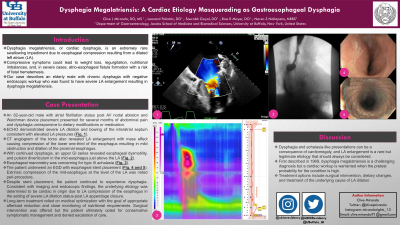
Introduction: Dysphagia megalatriensis, or cardiac dysphagia, is an extremely rare swallowing impediment due to esophageal compression resulting from a dilated left atrium (LA). Compressive symptoms could lead to weight loss, regurgitation, nutritional imbalances, and, in severe cases, atrio-esophageal fistula formation with a risk of fatal hematemesis. Our case describes an elderly male with chronic dysphagia with negative endoscopic workup who was found to have severe LA enlargement resulting in dysphagia megalatriensis.
Case Description/Methods: An 82-year-old male with atrial fibrillation status post AV nodal ablation and Watchman device placement presented for several months of abdominal pain and dysphagia unresponsive to dietary modifications or medication. Echocardiography demonstrated severe LA dilation and bowing of the interatrial septum consistent with elevated LA pressures (Fig. 1). A CT angiogram of the torso also revealed LA enlargement with mass effect causing compression of the lower one-third of the esophagus resulting in mild obstruction and dilation of the proximal esophagus. With continued dysphagia, an upper GI series revealed esophageal dysmotility, and pulsion diverticulum in the mid-esophagus just above the LA (Fig. 2). Esophageal manometry was concerning for type III achalasia (Fig. 3). The patient underwent an EGD with esophageal stent placement (Fig. 4). Extrinsic compression of the mid-esophagus at the level of the LA was noted peri-procedure. Despite stent placement, the patient continued to experience dysphagia. Consistent with imaging and endoscopic findings, the underlying etiology was determined to be cardiac in origin due to LA compression of the esophagus in the setting of severe LA dilation status-post LA appendage closure. Long-term treatment relied on medical optimization with the goal of appropriate afterload reduction and close monitoring of nutritional requirements. Surgical intervention was offered but the patient ultimately opted for conservative symptomatic management and denied escalation of care.
Discussion: Dysphagia and achalasia-like presentations can be a consequence of cardiomegaly, and LA enlargement is a rare but legitimate etiology that should always be considered. First described in 1969, dysphagia megalatriensis is a challenging diagnosis but a cardiac workup is warranted when the pretest probability for the condition is high. Treatment options include surgical intervention, dietary changes, and treatment of the underlying cause of LA dilation.

Disclosures:
Clive J. Miranda, DO, MS, Leonard Palatnic, DO, Sourabh Goyal, DO, Ross R. Moyer, DO, Naren S. Nallapeta, MBBS. P1901 - Dysphagia Megalatriensis: A Cardiac Etiology Masquerading as Gastroesophageal Dysphagia, ACG 2023 Annual Scientific Meeting Abstracts. Vancouver, BC, Canada: American College of Gastroenterology.
University at Buffalo, Buffalo, NY
Introduction: Dysphagia megalatriensis, or cardiac dysphagia, is an extremely rare swallowing impediment due to esophageal compression resulting from a dilated left atrium (LA). Compressive symptoms could lead to weight loss, regurgitation, nutritional imbalances, and, in severe cases, atrio-esophageal fistula formation with a risk of fatal hematemesis. Our case describes an elderly male with chronic dysphagia with negative endoscopic workup who was found to have severe LA enlargement resulting in dysphagia megalatriensis.
Case Description/Methods: An 82-year-old male with atrial fibrillation status post AV nodal ablation and Watchman device placement presented for several months of abdominal pain and dysphagia unresponsive to dietary modifications or medication. Echocardiography demonstrated severe LA dilation and bowing of the interatrial septum consistent with elevated LA pressures (Fig. 1). A CT angiogram of the torso also revealed LA enlargement with mass effect causing compression of the lower one-third of the esophagus resulting in mild obstruction and dilation of the proximal esophagus. With continued dysphagia, an upper GI series revealed esophageal dysmotility, and pulsion diverticulum in the mid-esophagus just above the LA (Fig. 2). Esophageal manometry was concerning for type III achalasia (Fig. 3). The patient underwent an EGD with esophageal stent placement (Fig. 4). Extrinsic compression of the mid-esophagus at the level of the LA was noted peri-procedure. Despite stent placement, the patient continued to experience dysphagia. Consistent with imaging and endoscopic findings, the underlying etiology was determined to be cardiac in origin due to LA compression of the esophagus in the setting of severe LA dilation status-post LA appendage closure. Long-term treatment relied on medical optimization with the goal of appropriate afterload reduction and close monitoring of nutritional requirements. Surgical intervention was offered but the patient ultimately opted for conservative symptomatic management and denied escalation of care.
Discussion: Dysphagia and achalasia-like presentations can be a consequence of cardiomegaly, and LA enlargement is a rare but legitimate etiology that should always be considered. First described in 1969, dysphagia megalatriensis is a challenging diagnosis but a cardiac workup is warranted when the pretest probability for the condition is high. Treatment options include surgical intervention, dietary changes, and treatment of the underlying cause of LA dilation.

Figure: Figure1: ECHO showing severe LA dilation and bowing of the interatrial septum consistent with elevated LA pressures.
Figure 2: Upper GI series concerning for esophageal dysmotility and mid-esophageal pulsion diverticulum just above the LA.
Figure 3: Esophageal manometry with readings concerning for type III achalasia.
Figure 4: EGD post-esophageal stent deployment.
Figure 2: Upper GI series concerning for esophageal dysmotility and mid-esophageal pulsion diverticulum just above the LA.
Figure 3: Esophageal manometry with readings concerning for type III achalasia.
Figure 4: EGD post-esophageal stent deployment.
Disclosures:
Clive Miranda indicated no relevant financial relationships.
Leonard Palatnic indicated no relevant financial relationships.
Sourabh Goyal indicated no relevant financial relationships.
Ross Moyer indicated no relevant financial relationships.
Naren Nallapeta indicated no relevant financial relationships.
Clive J. Miranda, DO, MS, Leonard Palatnic, DO, Sourabh Goyal, DO, Ross R. Moyer, DO, Naren S. Nallapeta, MBBS. P1901 - Dysphagia Megalatriensis: A Cardiac Etiology Masquerading as Gastroesophageal Dysphagia, ACG 2023 Annual Scientific Meeting Abstracts. Vancouver, BC, Canada: American College of Gastroenterology.
