Monday Poster Session
Category: Esophagus
P1909 - Lymphocytic Esophagitis: An Under-Recognized Cause of Dysphagia
Monday, October 23, 2023
10:30 AM - 4:15 PM PT
Location: Exhibit Hall

Has Audio
.jpg)
Anjella Manoharan, MS, MD
Rutgers New Jersey Medical School
Newark, NJ
Presenting Author(s)
Anjella Manoharan, MS, MD1, Vincent Wong, MD1, Dhanasekaran Ramasamy, MD2, Shiva Kumar, MD3
1Rutgers New Jersey Medical School, Newark, NJ; 2RWJ Barnabas Health, Livingston, NJ; 3Cleveland Clinic Abu Dhabi, Abu Dhabi, Abu Dhabi, United Arab Emirates
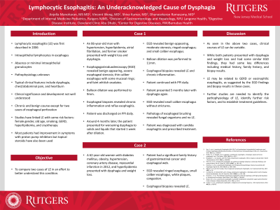
Introduction: Lymphocytic esophagitis (LE) is a rare entity characterized by esophageal intraepithelial lymphocytosis with evidence of epithelial injury and paucity of granulocytes. Its pathophysiology and natural history remain poorly defined. We report 2 cases of this rare entity, highlighting heterogeneity in its clinical presentation, endoscopic features, and response to treatment.
Case Description/Methods: An 86-year-old male, former smoker, presented with weight loss and dysphagia. Esophagogastroduodenoscopy (EGD) revealed a severe stenosis in the esophagus, narrow caliber esophagus with mucosal rings and whitish exudates. Biopsies from the proximal and distal esophagus revealed none to rare eosinophils (< 1 per high power field) and squamous mucosa with chronic inflammation. He underwent balloon dilation followed by acid suppressive therapy using proton pump inhibitors (PPIs) with symptomatic improvement. Repeat EGD with biopsy, which was performed 4 months later for recurrent dysphagia, revealed some improvement in endoscopic findings and increased intraepithelial lymphocytic infiltration on histology. Repeat balloon dilation with resumption of PPI therapy resulted in improvement of symptoms. Follow-up EGD with brushing 5 months later confirmed resolution of stricture and intraepithelial lymphocytosis.
A 62-year-old obese female with diabetes presented with progressive dysphagia and weight loss. EGD revealed ringed, small-caliber esophagus with small white plaques. Biopsies from the distal and proximal esophageal body were consistent with LE. PPI therapy was initiated with resolution of symptoms.
Discussion: Initially described in 2006, LE is a subset of chronic esophagitis with distinctive clinical and histologic features. Female preponderance has been noted and reported associations include pediatric inflammatory bowel disease, lichen planus, primary motility disorders, and smoking. The reported spectrum of clinical and endoscopic presentations is varied, with dysphagia being the predominant presentation. The overlapping symptoms and endoscopic features of LE and eosinophilic esophagitis (EoE) and responsiveness to acid suppressive therapy are noteworthy. Limited data exist regarding the natural history of LE and appropriate treatment plan, including the role of acid suppressive therapy. Whether LE is a distinct clinicopathological entity or a variable histologic manifestation of GERD or EoE needs further clarification.
Disclosures:
Anjella Manoharan, MS, MD1, Vincent Wong, MD1, Dhanasekaran Ramasamy, MD2, Shiva Kumar, MD3. P1909 - Lymphocytic Esophagitis: An Under-Recognized Cause of Dysphagia, ACG 2023 Annual Scientific Meeting Abstracts. Vancouver, BC, Canada: American College of Gastroenterology.
1Rutgers New Jersey Medical School, Newark, NJ; 2RWJ Barnabas Health, Livingston, NJ; 3Cleveland Clinic Abu Dhabi, Abu Dhabi, Abu Dhabi, United Arab Emirates
Introduction: Lymphocytic esophagitis (LE) is a rare entity characterized by esophageal intraepithelial lymphocytosis with evidence of epithelial injury and paucity of granulocytes. Its pathophysiology and natural history remain poorly defined. We report 2 cases of this rare entity, highlighting heterogeneity in its clinical presentation, endoscopic features, and response to treatment.
Case Description/Methods: An 86-year-old male, former smoker, presented with weight loss and dysphagia. Esophagogastroduodenoscopy (EGD) revealed a severe stenosis in the esophagus, narrow caliber esophagus with mucosal rings and whitish exudates. Biopsies from the proximal and distal esophagus revealed none to rare eosinophils (< 1 per high power field) and squamous mucosa with chronic inflammation. He underwent balloon dilation followed by acid suppressive therapy using proton pump inhibitors (PPIs) with symptomatic improvement. Repeat EGD with biopsy, which was performed 4 months later for recurrent dysphagia, revealed some improvement in endoscopic findings and increased intraepithelial lymphocytic infiltration on histology. Repeat balloon dilation with resumption of PPI therapy resulted in improvement of symptoms. Follow-up EGD with brushing 5 months later confirmed resolution of stricture and intraepithelial lymphocytosis.
A 62-year-old obese female with diabetes presented with progressive dysphagia and weight loss. EGD revealed ringed, small-caliber esophagus with small white plaques. Biopsies from the distal and proximal esophageal body were consistent with LE. PPI therapy was initiated with resolution of symptoms.
Discussion: Initially described in 2006, LE is a subset of chronic esophagitis with distinctive clinical and histologic features. Female preponderance has been noted and reported associations include pediatric inflammatory bowel disease, lichen planus, primary motility disorders, and smoking. The reported spectrum of clinical and endoscopic presentations is varied, with dysphagia being the predominant presentation. The overlapping symptoms and endoscopic features of LE and eosinophilic esophagitis (EoE) and responsiveness to acid suppressive therapy are noteworthy. Limited data exist regarding the natural history of LE and appropriate treatment plan, including the role of acid suppressive therapy. Whether LE is a distinct clinicopathological entity or a variable histologic manifestation of GERD or EoE needs further clarification.
Disclosures:
Anjella Manoharan indicated no relevant financial relationships.
Vincent Wong indicated no relevant financial relationships.
Dhanasekaran Ramasamy indicated no relevant financial relationships.
Shiva Kumar indicated no relevant financial relationships.
Anjella Manoharan, MS, MD1, Vincent Wong, MD1, Dhanasekaran Ramasamy, MD2, Shiva Kumar, MD3. P1909 - Lymphocytic Esophagitis: An Under-Recognized Cause of Dysphagia, ACG 2023 Annual Scientific Meeting Abstracts. Vancouver, BC, Canada: American College of Gastroenterology.
