Monday Poster Session
Category: Colon
P1713 - A Diagnostic Dilemma: Sigmoid Leiomyoma Masked as a Colon Polyp
Monday, October 23, 2023
10:30 AM - 4:15 PM PT
Location: Exhibit Hall

Has Audio

Keshavi Mahesh, MD
Broward Health North
Deerfield Beach, FL
Presenting Author(s)
Keshavi Mahesh, MD1, Aryama Sharma, MD2
1Broward Health North, Deerfield Beach, FL; 2Broward Health Medical Center, Fort Lauderdale, FL
Introduction: Sigmoid leiomyoma, a rare benign tumor of the colon, accounts for only 3% of gastrointestinal smooth muscle tumors. This condition is predominantly observed in males, with the average age of detection at 62 years. Smooth muscle neoplasms can arise from either the muscularis mucosae or the proper muscle layer, resulting in the formation of polypoid or intramural lesions. Polypoid leiomyomas are frequently found in the rectosigmoid area and can be effectively treated through endoscopic resection. On the other hand, local surgical excision is the preferred treatment for intramural leiomyomas. Although most leiomyomas are benign it is important to pay attention to the size, location, and histological appearance as even benign looking smooth muscle tumors may metastasize. In this case we present a rare case of sigmoid leiomyoma disguised as a colonic polyp.
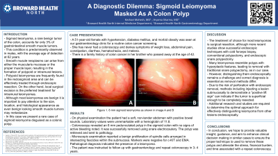
Case Description/Methods: A 51-year-old female with hypertension, diabetes mellitus, and obesity was seen at our gastroenterology clinic for routine colon cancer screening. She has never had a colonoscopy and denies symptoms of weight loss, abdominal pain, constipation, diarrhea, hematochezia, and melena. There is a family history of colon cancer in her brother who passed away at the age of 42. Physical examination and laboratory values were unremarkable. Colonoscopy revealed an 8 mm polyp in the sigmoid colon which was successfully removed using snare cautery. Surgical pathology revealed the sigmoid polyp to be a submucosal leiomyoma negative for c-KIT and epithelial membrane antigen (EMA). The patient was instructed to follow up with gastroenterology and repeat colonoscopy in 3- 4 years.
Discussion: The treatment of choice for most leiomyomas is surgical resection, although more recent studies show successful endoscopic techniques with cold forceps biopsy, conventional polypectomy, and endoscopic snare polypectomy. Many leiomyomas resemble polyps with hyperplastic features, leading to removal with traditional snare polypectomy, as in our case. However, distinguishing them endoscopically remains a challenge and correct diagnosis is essential as removal methods differ. Additional research is required to determine the optimal approach for effectively distinguishing leiomyoma from other lesions endoscopically. In conclusion, we hope to provide valuable insight and guidance and aim to enhance clinical decision making in similar cases to alleviate the stress, financial burden, and time associated with a repeat colonoscopy.

Disclosures:
Keshavi Mahesh, MD1, Aryama Sharma, MD2. P1713 - A Diagnostic Dilemma: Sigmoid Leiomyoma Masked as a Colon Polyp, ACG 2023 Annual Scientific Meeting Abstracts. Vancouver, BC, Canada: American College of Gastroenterology.
1Broward Health North, Deerfield Beach, FL; 2Broward Health Medical Center, Fort Lauderdale, FL
Introduction: Sigmoid leiomyoma, a rare benign tumor of the colon, accounts for only 3% of gastrointestinal smooth muscle tumors. This condition is predominantly observed in males, with the average age of detection at 62 years. Smooth muscle neoplasms can arise from either the muscularis mucosae or the proper muscle layer, resulting in the formation of polypoid or intramural lesions. Polypoid leiomyomas are frequently found in the rectosigmoid area and can be effectively treated through endoscopic resection. On the other hand, local surgical excision is the preferred treatment for intramural leiomyomas. Although most leiomyomas are benign it is important to pay attention to the size, location, and histological appearance as even benign looking smooth muscle tumors may metastasize. In this case we present a rare case of sigmoid leiomyoma disguised as a colonic polyp.
Case Description/Methods: A 51-year-old female with hypertension, diabetes mellitus, and obesity was seen at our gastroenterology clinic for routine colon cancer screening. She has never had a colonoscopy and denies symptoms of weight loss, abdominal pain, constipation, diarrhea, hematochezia, and melena. There is a family history of colon cancer in her brother who passed away at the age of 42. Physical examination and laboratory values were unremarkable. Colonoscopy revealed an 8 mm polyp in the sigmoid colon which was successfully removed using snare cautery. Surgical pathology revealed the sigmoid polyp to be a submucosal leiomyoma negative for c-KIT and epithelial membrane antigen (EMA). The patient was instructed to follow up with gastroenterology and repeat colonoscopy in 3- 4 years.
Discussion: The treatment of choice for most leiomyomas is surgical resection, although more recent studies show successful endoscopic techniques with cold forceps biopsy, conventional polypectomy, and endoscopic snare polypectomy. Many leiomyomas resemble polyps with hyperplastic features, leading to removal with traditional snare polypectomy, as in our case. However, distinguishing them endoscopically remains a challenge and correct diagnosis is essential as removal methods differ. Additional research is required to determine the optimal approach for effectively distinguishing leiomyoma from other lesions endoscopically. In conclusion, we hope to provide valuable insight and guidance and aim to enhance clinical decision making in similar cases to alleviate the stress, financial burden, and time associated with a repeat colonoscopy.

Figure: Figure 1. 8 mm sigmoid leiomyoma visualized during the colonoscopy with characteristics similar to a hyperplastic polyp.
Disclosures:
Keshavi Mahesh indicated no relevant financial relationships.
Aryama Sharma indicated no relevant financial relationships.
Keshavi Mahesh, MD1, Aryama Sharma, MD2. P1713 - A Diagnostic Dilemma: Sigmoid Leiomyoma Masked as a Colon Polyp, ACG 2023 Annual Scientific Meeting Abstracts. Vancouver, BC, Canada: American College of Gastroenterology.
