Monday Poster Session
Category: Colon
P1717 - Disseminated Invasive Lobular Carcinoma Metastasis of the Gastrointestinal Tract: An Oncologic Anomaly
Monday, October 23, 2023
10:30 AM - 4:15 PM PT
Location: Exhibit Hall

Has Audio

Elliott Draughn
Edward Via College of Osteopathic Medicine
Roanoke, Virginia
Presenting Author(s)
Elliott Draughn, 1, Karri Vandana, MD2, Chuan Long Miao, MD, FACG2
1Edward Via College of Osteopathic Medicine, Roanoke, VA; 2Lewis Gale Medical Center, Roanoke, VA
Introduction: Breast malignancies have a known propensity for metastasis to bone, lung, and liver. However, gastrointestinal metastatic disease is a rare phenomenon from primary breast malignancy. We present a rare case of invasive lobular carcinoma with extensive metastasis throughout the GI tract, a relapse manifesting 19 years after initial diagnosis of primary breast cancer diagnosis.
Case Description/Methods: A 55-year-old female with a medical history of right breast cancer status post bilateral mastectomy in 2003, presented with 4 weeks of nausea, vomiting, and dull abdominal pain. Pathology revealed poorly differentiated carcinoma consistent with metastatic lobular carcinoma of primary breast tissue. Tumor marker CA 27-29 was significantly elevated at 436.6 U/mL. CT revealed bowel wall thickening and small bowel obstruction. Endoscopy demonstrated multiple stenoses and scattered granular, nodular, and erythematous, cobblestone appearing mucosa throughout the GI tract. Severe stenoses at pylorus and multiple colon locations required ultra-thin pediatric endoscope to traverse. Pathology demonstrated metastatic invasive lobular carcinoma involving the esophagus, stomach, duodenum, colon, and rectum. Tumors exhibited strong ER positivity, weak PR positivity, and low HER2 expressivity. An adjuvant therapy of Letrozole + Ribociclib was immediately initiated, and patient was discharged to follow up with oncology. PET-CT demonstrated extensive multi-system metastasis, which revealed increased uptake by the brain parenchyma, liver, spleen, stomach, small bowel, colon, rectum, axial and appendicular skeleton. Due to presence of skeletal metastasis, Denosumab was added for suppression of skeletal metastasis and secondary fracture prevention. The patient had resolution of GI symptoms, with reduction in skin metastasis lesion size at oncology follow up visits.
Discussion: Only 0.6% of all reported primary breast cancers involve GI spread [1]. Database studies reported a median time of breast cancer recurrence as GI metastases was 9.62 years after diagnosis of primary cancer [2]. In our case, the diagnosis was made 19 years after diagnosis of her primary breast cancer. To our knowledge, there are currently no cases reporting this extensiveness of metastasis throughout the GI tract following nearly two decades of clinical breast cancer remission. The unprecedented degree of disseminated metastasis and tumor burden posed significant challenge for the validation of predictive prognosis and therapy response.

Disclosures:
Elliott Draughn, 1, Karri Vandana, MD2, Chuan Long Miao, MD, FACG2. P1717 - Disseminated Invasive Lobular Carcinoma Metastasis of the Gastrointestinal Tract: An Oncologic Anomaly, ACG 2023 Annual Scientific Meeting Abstracts. Vancouver, BC, Canada: American College of Gastroenterology.
1Edward Via College of Osteopathic Medicine, Roanoke, VA; 2Lewis Gale Medical Center, Roanoke, VA
Introduction: Breast malignancies have a known propensity for metastasis to bone, lung, and liver. However, gastrointestinal metastatic disease is a rare phenomenon from primary breast malignancy. We present a rare case of invasive lobular carcinoma with extensive metastasis throughout the GI tract, a relapse manifesting 19 years after initial diagnosis of primary breast cancer diagnosis.
Case Description/Methods: A 55-year-old female with a medical history of right breast cancer status post bilateral mastectomy in 2003, presented with 4 weeks of nausea, vomiting, and dull abdominal pain. Pathology revealed poorly differentiated carcinoma consistent with metastatic lobular carcinoma of primary breast tissue. Tumor marker CA 27-29 was significantly elevated at 436.6 U/mL. CT revealed bowel wall thickening and small bowel obstruction. Endoscopy demonstrated multiple stenoses and scattered granular, nodular, and erythematous, cobblestone appearing mucosa throughout the GI tract. Severe stenoses at pylorus and multiple colon locations required ultra-thin pediatric endoscope to traverse. Pathology demonstrated metastatic invasive lobular carcinoma involving the esophagus, stomach, duodenum, colon, and rectum. Tumors exhibited strong ER positivity, weak PR positivity, and low HER2 expressivity. An adjuvant therapy of Letrozole + Ribociclib was immediately initiated, and patient was discharged to follow up with oncology. PET-CT demonstrated extensive multi-system metastasis, which revealed increased uptake by the brain parenchyma, liver, spleen, stomach, small bowel, colon, rectum, axial and appendicular skeleton. Due to presence of skeletal metastasis, Denosumab was added for suppression of skeletal metastasis and secondary fracture prevention. The patient had resolution of GI symptoms, with reduction in skin metastasis lesion size at oncology follow up visits.
Discussion: Only 0.6% of all reported primary breast cancers involve GI spread [1]. Database studies reported a median time of breast cancer recurrence as GI metastases was 9.62 years after diagnosis of primary cancer [2]. In our case, the diagnosis was made 19 years after diagnosis of her primary breast cancer. To our knowledge, there are currently no cases reporting this extensiveness of metastasis throughout the GI tract following nearly two decades of clinical breast cancer remission. The unprecedented degree of disseminated metastasis and tumor burden posed significant challenge for the validation of predictive prognosis and therapy response.

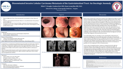
Figure: Endoscopy and Colonoscopy evaluation of lesions: Endoscopy demonstrated Schatzki ring at the gastroesophageal junction (a), scattered inflammation and erythema in stomach (b), and severe intrinsic stenosis of pylorus (c). Colonoscopy demonstrating intrinsic stenosis of proximal rectum (d), and nodular erythematous mucosa of the transverse colon (e) and cecum (f).
Disclosures:
Elliott Draughn indicated no relevant financial relationships.
Karri Vandana indicated no relevant financial relationships.
Chuan Long Miao indicated no relevant financial relationships.
Elliott Draughn, 1, Karri Vandana, MD2, Chuan Long Miao, MD, FACG2. P1717 - Disseminated Invasive Lobular Carcinoma Metastasis of the Gastrointestinal Tract: An Oncologic Anomaly, ACG 2023 Annual Scientific Meeting Abstracts. Vancouver, BC, Canada: American College of Gastroenterology.
