Monday Poster Session
Category: Colon
P1731 - A Rare Case of Heterotopic Ileocecal Tissue Found at the Splenic Flexure
Monday, October 23, 2023
10:30 AM - 4:15 PM PT
Location: Exhibit Hall

Has Audio

Megan B. Ghai, MD, MPH, MA
University of Arizona College of Medicine
Phoenix, AZ
Presenting Author(s)
Megan B. Ghai, MD, MPH, MA1, Natasha Narang, DO1, Donald Tschirhart, MD2, Natalee Campbell, MD3
1University of Arizona College of Medicine, Phoenix, AZ; 2VA Medical Center Phoenix, Phoenix, AZ; 3Phoenix VA Health Care System, Phoenix, AZ
Introduction: While gastric heterotopia is a known condition in which normal gastric tissue is found an unexpected sites, there have been no reports of small intestinal heterotopia. Presented is a rare case of heterotopic ileocecal tissue found at the splenic flexure.
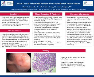
Case Description/Methods: A 62-year-old male with a past medical history of irritable bowel syndrome, type two diabetes mellitus, testicular cancer, and pulmonary embolism on anticoagulation presented to the hospital with dizziness, abdominal pain, and nausea. He reportedly had three days of black stools that had resolved prior to presentation. He was hemodynamically stable but blood work revealed a hemoglobin level of 10.2 g/dL from a baseline of 14 g/dL. Esophagogastroduodenoscopy was performed that showed no evidence of peptic ulcer disease, esophagitis, or strictures. Colonoscopy demonstrated mucosal pallor throughout the colon but no sources of bleeding. Diverticulosis in the descending and sigmoid colon was also noted. Finally, a friable raised lesion was identified at the splenic flexure and was biopsied at 70 cm (Figure 1a). No other biopsies were taken. Final histology demonstrated tissue consistent with the ileocecal valve (Figure 1b).
Discussion: There have been no reported cases of heterotopic ileocecal tissue found in the colon. While mislabeled biopsy samples would be the most plausible explanation, the only biopsy taken in this case was of the friable mucosa seen at the splenic flexure. While the clinical significance of ileocecal heterotopia is unknown, it warrants further evaluation as gastric heterotopia can be associated with malignant transformation. Further, gastric heterotopia can be associated with abdominal pain, nausea, and bleeding which could explain the patient’s presenting symptoms as no explanation for his hemoglobin drop was identified on esophagogastroduodenoscopy or colonoscopy.

Disclosures:
Megan B. Ghai, MD, MPH, MA1, Natasha Narang, DO1, Donald Tschirhart, MD2, Natalee Campbell, MD3. P1731 - A Rare Case of Heterotopic Ileocecal Tissue Found at the Splenic Flexure, ACG 2023 Annual Scientific Meeting Abstracts. Vancouver, BC, Canada: American College of Gastroenterology.
1University of Arizona College of Medicine, Phoenix, AZ; 2VA Medical Center Phoenix, Phoenix, AZ; 3Phoenix VA Health Care System, Phoenix, AZ
Introduction: While gastric heterotopia is a known condition in which normal gastric tissue is found an unexpected sites, there have been no reports of small intestinal heterotopia. Presented is a rare case of heterotopic ileocecal tissue found at the splenic flexure.
Case Description/Methods: A 62-year-old male with a past medical history of irritable bowel syndrome, type two diabetes mellitus, testicular cancer, and pulmonary embolism on anticoagulation presented to the hospital with dizziness, abdominal pain, and nausea. He reportedly had three days of black stools that had resolved prior to presentation. He was hemodynamically stable but blood work revealed a hemoglobin level of 10.2 g/dL from a baseline of 14 g/dL. Esophagogastroduodenoscopy was performed that showed no evidence of peptic ulcer disease, esophagitis, or strictures. Colonoscopy demonstrated mucosal pallor throughout the colon but no sources of bleeding. Diverticulosis in the descending and sigmoid colon was also noted. Finally, a friable raised lesion was identified at the splenic flexure and was biopsied at 70 cm (Figure 1a). No other biopsies were taken. Final histology demonstrated tissue consistent with the ileocecal valve (Figure 1b).
Discussion: There have been no reported cases of heterotopic ileocecal tissue found in the colon. While mislabeled biopsy samples would be the most plausible explanation, the only biopsy taken in this case was of the friable mucosa seen at the splenic flexure. While the clinical significance of ileocecal heterotopia is unknown, it warrants further evaluation as gastric heterotopia can be associated with malignant transformation. Further, gastric heterotopia can be associated with abdominal pain, nausea, and bleeding which could explain the patient’s presenting symptoms as no explanation for his hemoglobin drop was identified on esophagogastroduodenoscopy or colonoscopy.

Figure: Figure 1a: Friable tissue seen at the splenic flexure (see arrow).
Figure 1b: Low-power photomicrograph, 40X, H&E stain. Coexistence of small intestinal and colonic-type mucosa at the splenic flexure.
Figure 1b: Low-power photomicrograph, 40X, H&E stain. Coexistence of small intestinal and colonic-type mucosa at the splenic flexure.
Disclosures:
Megan Ghai indicated no relevant financial relationships.
Natasha Narang indicated no relevant financial relationships.
Donald Tschirhart indicated no relevant financial relationships.
Natalee Campbell indicated no relevant financial relationships.
Megan B. Ghai, MD, MPH, MA1, Natasha Narang, DO1, Donald Tschirhart, MD2, Natalee Campbell, MD3. P1731 - A Rare Case of Heterotopic Ileocecal Tissue Found at the Splenic Flexure, ACG 2023 Annual Scientific Meeting Abstracts. Vancouver, BC, Canada: American College of Gastroenterology.
