Monday Poster Session
Category: Biliary/Pancreas
P1500 - Metastatic Testicular Tumors Masquerading as Pancreatic Cancer: A Case Series
Monday, October 23, 2023
10:30 AM - 4:15 PM PT
Location: Exhibit Hall

Has Audio

Ammar Qureshi, MD
University of California Riverside School of Medicine
Riverside, CA
Presenting Author(s)
Mahtab Naji, MD1, Ammar Qureshi, MD1, Harold Paredes, MD2
1University of California Riverside School of Medicine, Riverside, CA; 2University of California Riverside, Riverside, CA
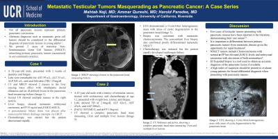
Introduction: Not all pancreatic lesions represent primary pancreatic carcinomas. Alternate diagnoses such as metastatic germ cell tumors should be considered in the differential diagnosis of pancreatic lesions in young adults. We present 2 cases of testicular Non-Seminomatous Germ Cell Tumors (NSGCT) mimicking primary pancreatic tumors encountered in our community practice.
Case Description/Methods: Case 1:
A 28-year-old male, presented with 2 weeks of jaundice and fatigue. Labs were remarkable for AST 59u/L, ALT 53u/L, ALP 845u/L, and total bilirubin (TB) >25mg/dl. CT and MRCP showed 2 masses in the liver causing mass effect with intrahepatic ductal dilatation and an ill-defined lesion in the pancreatic head measuring 6x8cm (Image 1). Scrotal US showed multiple masses in the right testicle. A liver biopsy showed metastatic embryonal carcinoma. 𝞪-FP 35ng/ml and 𝛽-HCG 66IU/L. A percutaneous biliary drain was placed after unsuccessful biliary drainage attempts via ERCP. Chemotherapy was started but the patient deteriorated rapidly.
Case 2:
A 43-year-old male with a history of testicular cancer, treated with orchiectomy and chemotherapy at age 12, presented with weight loss, icterus, and fatigue. Labs showed TB of 2.4mg/dl, ALT 42u/L, AST 45u/L, and ALP 280u/L. 𝛽-hCG 105526IU/L and 𝞪-FP 1.9ng/ml. CT showed a complex pancreatic head mass measuring 13cm and multiple liver lesions (Image 2). EUS demonstrated a 11cmx10cm heterogeneous mass with areas of cystic degeneration in the pancreatic head (Image 3). Biopsy was consistent with metastatic choriocarcinoma. The concomitant liver biopsy also confirmed poorly differentiated metastatic NSGCT. Chemotherapy was initiated but the patient rapidly developed multiorgan failure.
Discussion: Few cases of testicular tumors presenting with pancreatic masses have been reported in the literature, demonstrating their rare nature. It is important to differentiate between primary pancreatic tumors from metastatic disease given the opportunity for rapid treatment. NSGCT tumors include Choriocarcinoma with normal 𝞪-FP but elevated 𝛽-HCG levels and embryonal carcinoma with elevations of both seromarkers. EUS-guided biopsy is a tool used to obtain an accurate diagnosis of the pancreatic lesion if available. A high index of suspicion should be present to evaluate young patients for broad differential diagnosis when presenting with pancreatic masses.

Disclosures:
Mahtab Naji, MD1, Ammar Qureshi, MD1, Harold Paredes, MD2. P1500 - Metastatic Testicular Tumors Masquerading as Pancreatic Cancer: A Case Series, ACG 2023 Annual Scientific Meeting Abstracts. Vancouver, BC, Canada: American College of Gastroenterology.
1University of California Riverside School of Medicine, Riverside, CA; 2University of California Riverside, Riverside, CA
Introduction: Not all pancreatic lesions represent primary pancreatic carcinomas. Alternate diagnoses such as metastatic germ cell tumors should be considered in the differential diagnosis of pancreatic lesions in young adults. We present 2 cases of testicular Non-Seminomatous Germ Cell Tumors (NSGCT) mimicking primary pancreatic tumors encountered in our community practice.
Case Description/Methods: Case 1:
A 28-year-old male, presented with 2 weeks of jaundice and fatigue. Labs were remarkable for AST 59u/L, ALT 53u/L, ALP 845u/L, and total bilirubin (TB) >25mg/dl. CT and MRCP showed 2 masses in the liver causing mass effect with intrahepatic ductal dilatation and an ill-defined lesion in the pancreatic head measuring 6x8cm (Image 1). Scrotal US showed multiple masses in the right testicle. A liver biopsy showed metastatic embryonal carcinoma. 𝞪-FP 35ng/ml and 𝛽-HCG 66IU/L. A percutaneous biliary drain was placed after unsuccessful biliary drainage attempts via ERCP. Chemotherapy was started but the patient deteriorated rapidly.
Case 2:
A 43-year-old male with a history of testicular cancer, treated with orchiectomy and chemotherapy at age 12, presented with weight loss, icterus, and fatigue. Labs showed TB of 2.4mg/dl, ALT 42u/L, AST 45u/L, and ALP 280u/L. 𝛽-hCG 105526IU/L and 𝞪-FP 1.9ng/ml. CT showed a complex pancreatic head mass measuring 13cm and multiple liver lesions (Image 2). EUS demonstrated a 11cmx10cm heterogeneous mass with areas of cystic degeneration in the pancreatic head (Image 3). Biopsy was consistent with metastatic choriocarcinoma. The concomitant liver biopsy also confirmed poorly differentiated metastatic NSGCT. Chemotherapy was initiated but the patient rapidly developed multiorgan failure.
Discussion: Few cases of testicular tumors presenting with pancreatic masses have been reported in the literature, demonstrating their rare nature. It is important to differentiate between primary pancreatic tumors from metastatic disease given the opportunity for rapid treatment. NSGCT tumors include Choriocarcinoma with normal 𝞪-FP but elevated 𝛽-HCG levels and embryonal carcinoma with elevations of both seromarkers. EUS-guided biopsy is a tool used to obtain an accurate diagnosis of the pancreatic lesion if available. A high index of suspicion should be present to evaluate young patients for broad differential diagnosis when presenting with pancreatic masses.

Figure: Image 1: MRCP showing a lesion in the pancreatic head measuring 6x8cm
Image 2: CT Abdomen and pelvis, showing a complex pancreatic head mass measuring 13cm and multiple liver lesions
Image 3: EUS showing a 11cmx10cm heterogeneous mass with areas of cystic degeneration in the
pancreatic head
Image 2: CT Abdomen and pelvis, showing a complex pancreatic head mass measuring 13cm and multiple liver lesions
Image 3: EUS showing a 11cmx10cm heterogeneous mass with areas of cystic degeneration in the
pancreatic head
Disclosures:
Mahtab Naji indicated no relevant financial relationships.
Ammar Qureshi indicated no relevant financial relationships.
Harold Paredes indicated no relevant financial relationships.
Mahtab Naji, MD1, Ammar Qureshi, MD1, Harold Paredes, MD2. P1500 - Metastatic Testicular Tumors Masquerading as Pancreatic Cancer: A Case Series, ACG 2023 Annual Scientific Meeting Abstracts. Vancouver, BC, Canada: American College of Gastroenterology.
