Tuesday Poster Session
Category: Interventional Endoscopy
P3739 - Bouveret Syndrome: The Absolute Gallstone
Tuesday, October 24, 2023
10:30 AM - 4:00 PM PT
Location: Exhibit Hall

Has Audio

Taimur S. Muzammil, MD
Allegheny Health Network
Pittsburgh, PA
Presenting Author(s)
Taimur S. Muzammil, MD1, Muhammad Ali Butt, MD2
1Allegheny Health Network, Pittsburgh, PA; 2Medicine Institute, Allegheny Health Network, Pittsburgh, PA
Introduction: Bouveret syndrome is an impaction of a large gallstone within the pyloric channel that usually requires surgery to remove the impacted stone, repair the fistula, and remove the gallbladder. We present a case of a patient with a gallstone greater than 5cm with conditions not allowing for surgical interventions initially leading to endoscopic attempt at removal.
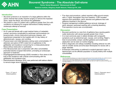
Case Description/Methods: An 81-year-old female with a past medical history of metastatic ovarian carcinoma complicated by peritoneal carcinomatosis (on bevacizumab), chronic obstructive pulmonary disease, and hypertension presented to the hospital with 3-4 day history of abdominal pain, nausea, and vomiting. Physical exam and initial laboratory workup were unremarkable. CT abdomen and pelvis (CTAP) noted 2x3 cm gallstones in the gallbladder and duodenum concerning for a cholecystoduodenal fistula without obstruction or perforation. Prior Bevacizumab use in conjunction with other comorbidities precluded elective surgical intervention, prompting an endoscopic approach to treatment. Esophagogastroduodenoscopy (EGD) revealed a >5cm stone in the bulb of the duodenum with a fistula communicating with the gallbladder and duodenal ulcer. Electrohydraulic lithotripsy (EHL) was performed with balloon dilation to remove larger stone fragments. Five days post-procedure, patient reported coffee ground emesis with a 1.4g/dL hemoglobin drop from baseline. CTAP revealed sigmoid colon perforation, pelvic abscess and gallstone ileus with gallstone impacted in the jejunum. Surgical management entailed gallstone removal, drainage of pelvic abscess, peritoneal washing with a diverting ileostomy. Post-operative course was uncomplicated.
Discussion: Bouveret syndrome is a rare form of gallstone ileus causing gastric outlet obstruction with stones typically greater than 2.5cm . It is most commonly seen in elderly females with nonspecific symptoms such as abdominal pain, nausea, and vomiting. Rigler’s triad of pneumobilia, ectopic calcified gallstone and small bowel obstruction can be seen on routine imaging. EGD is diagnostic and utilizes modalities including retrieval via roth-nets for smaller stones and EHL/laser lithotripsies for stones with a larger diameter. Endoscopic intervention is preferred to surgical approach (open vs laparoscopic) as the afflicted population is usually elderly with multiple comorbidities.

Disclosures:
Taimur S. Muzammil, MD1, Muhammad Ali Butt, MD2. P3739 - Bouveret Syndrome: The Absolute Gallstone, ACG 2023 Annual Scientific Meeting Abstracts. Vancouver, BC, Canada: American College of Gastroenterology.
1Allegheny Health Network, Pittsburgh, PA; 2Medicine Institute, Allegheny Health Network, Pittsburgh, PA
Introduction: Bouveret syndrome is an impaction of a large gallstone within the pyloric channel that usually requires surgery to remove the impacted stone, repair the fistula, and remove the gallbladder. We present a case of a patient with a gallstone greater than 5cm with conditions not allowing for surgical interventions initially leading to endoscopic attempt at removal.
Case Description/Methods: An 81-year-old female with a past medical history of metastatic ovarian carcinoma complicated by peritoneal carcinomatosis (on bevacizumab), chronic obstructive pulmonary disease, and hypertension presented to the hospital with 3-4 day history of abdominal pain, nausea, and vomiting. Physical exam and initial laboratory workup were unremarkable. CT abdomen and pelvis (CTAP) noted 2x3 cm gallstones in the gallbladder and duodenum concerning for a cholecystoduodenal fistula without obstruction or perforation. Prior Bevacizumab use in conjunction with other comorbidities precluded elective surgical intervention, prompting an endoscopic approach to treatment. Esophagogastroduodenoscopy (EGD) revealed a >5cm stone in the bulb of the duodenum with a fistula communicating with the gallbladder and duodenal ulcer. Electrohydraulic lithotripsy (EHL) was performed with balloon dilation to remove larger stone fragments. Five days post-procedure, patient reported coffee ground emesis with a 1.4g/dL hemoglobin drop from baseline. CTAP revealed sigmoid colon perforation, pelvic abscess and gallstone ileus with gallstone impacted in the jejunum. Surgical management entailed gallstone removal, drainage of pelvic abscess, peritoneal washing with a diverting ileostomy. Post-operative course was uncomplicated.
Discussion: Bouveret syndrome is a rare form of gallstone ileus causing gastric outlet obstruction with stones typically greater than 2.5cm . It is most commonly seen in elderly females with nonspecific symptoms such as abdominal pain, nausea, and vomiting. Rigler’s triad of pneumobilia, ectopic calcified gallstone and small bowel obstruction can be seen on routine imaging. EGD is diagnostic and utilizes modalities including retrieval via roth-nets for smaller stones and EHL/laser lithotripsies for stones with a larger diameter. Endoscopic intervention is preferred to surgical approach (open vs laparoscopic) as the afflicted population is usually elderly with multiple comorbidities.

Figure: CT and endoscopic visualization of gallstone.
Disclosures:
Taimur Muzammil indicated no relevant financial relationships.
Muhammad Ali Butt indicated no relevant financial relationships.
Taimur S. Muzammil, MD1, Muhammad Ali Butt, MD2. P3739 - Bouveret Syndrome: The Absolute Gallstone, ACG 2023 Annual Scientific Meeting Abstracts. Vancouver, BC, Canada: American College of Gastroenterology.
