Tuesday Poster Session
Category: Interventional Endoscopy
P3748 - Gallbladder Necrosum
Tuesday, October 24, 2023
10:30 AM - 4:00 PM PT
Location: Exhibit Hall


Natalie Wilson, MD
University of Minnesota
Minneapolis, MN
Presenting Author(s)
Natalie Wilson, MD1, Vijay S. Are, MD1, Mohammad Bilal, MD2
1University of Minnesota, Minneapolis, MN; 2Minneapolis VA Medical Center, Minneapolis, MN
Introduction: Endoscopic ultrasound-guided gallbladder drainage (EUS-GBD) using lumen-apposing metal stents (LAMS) has allowed for safe and effective endoscopic management of acute cholecystitis in nonsurgical candidates. Despite increasing adoption of EUS-GBD, aspects of this technique are still evolving. Here, we describe a case of acute necrotizing cholecystitis which was successfully managed with removal of large piece of necrotic gallbladder tissue using a LAMS.
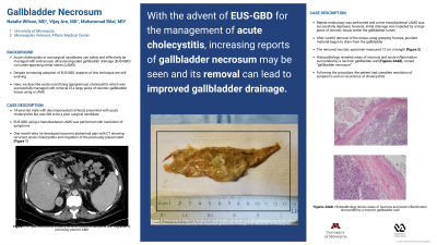
Case Description/Methods: A 74-year-old male with decompensated cirrhosis presented with acute cholecystitis. He was deemed a poor surgical candidate and therefore EUS-GBD using a transduodenal LAMS was performed. The patient’s symptoms resolved until one month later when he developed recurrent abdominal pain with imaging showing acute recurrent cholecystitis and migration of the previously placed stent [Figure 1A]. Repeat endoscopy was performed and a new transduodenal LAMS was successfully deployed, however, initial drainage was impeded by a large piece of necrotic tissue within the gallbladder lumen. After careful removal of the tissue using grasping forceps, purulent material began to drain from the gallbladder. The removed necrotic specimen measured 12 cm in length [Figure 1B]. Histopathology revealed areas of necrosis and acute inflammation surrounded by a necrotic gallbladder wall [Figure 1C, 1D], coined “gallbladder necrosum”. Following the procedure, the patient had complete resolution of symptoms and no recurrence of cholecystitis.
Discussion: With the advent of EUS-GBD for the management of acute cholecystitis, increasing reports of gallbladder necrosum may be seen and its removal can lead to improved gallbladder drainage.

Disclosures:
Natalie Wilson, MD1, Vijay S. Are, MD1, Mohammad Bilal, MD2. P3748 - Gallbladder Necrosum, ACG 2023 Annual Scientific Meeting Abstracts. Vancouver, BC, Canada: American College of Gastroenterology.
1University of Minnesota, Minneapolis, MN; 2Minneapolis VA Medical Center, Minneapolis, MN
Introduction: Endoscopic ultrasound-guided gallbladder drainage (EUS-GBD) using lumen-apposing metal stents (LAMS) has allowed for safe and effective endoscopic management of acute cholecystitis in nonsurgical candidates. Despite increasing adoption of EUS-GBD, aspects of this technique are still evolving. Here, we describe a case of acute necrotizing cholecystitis which was successfully managed with removal of large piece of necrotic gallbladder tissue using a LAMS.
Case Description/Methods: A 74-year-old male with decompensated cirrhosis presented with acute cholecystitis. He was deemed a poor surgical candidate and therefore EUS-GBD using a transduodenal LAMS was performed. The patient’s symptoms resolved until one month later when he developed recurrent abdominal pain with imaging showing acute recurrent cholecystitis and migration of the previously placed stent [Figure 1A]. Repeat endoscopy was performed and a new transduodenal LAMS was successfully deployed, however, initial drainage was impeded by a large piece of necrotic tissue within the gallbladder lumen. After careful removal of the tissue using grasping forceps, purulent material began to drain from the gallbladder. The removed necrotic specimen measured 12 cm in length [Figure 1B]. Histopathology revealed areas of necrosis and acute inflammation surrounded by a necrotic gallbladder wall [Figure 1C, 1D], coined “gallbladder necrosum”. Following the procedure, the patient had complete resolution of symptoms and no recurrence of cholecystitis.
Discussion: With the advent of EUS-GBD for the management of acute cholecystitis, increasing reports of gallbladder necrosum may be seen and its removal can lead to improved gallbladder drainage.

Figure: Figure 1. A) CT imaging of the abdomen shows gallbladder inflammation, wall thickening, and gallstones consistent with recurrent cholecystitis. The previously placed lumen apposing metal stent has migrated and can no longer be seen. B) Photograph of the 12 cm necrotic gallbladder specimen, coined gallbladder necrosum, which was removed endoscopically with improved gallbladder drainage and resolution of cholecystitis. C, D). Histopathology of the resected specimen shows a necrotic gallbladder wall with areas of necrosis and acute inflammation.
Disclosures:
Natalie Wilson indicated no relevant financial relationships.
Vijay Are indicated no relevant financial relationships.
Mohammad Bilal: Boston Scientific – Consultant.
Natalie Wilson, MD1, Vijay S. Are, MD1, Mohammad Bilal, MD2. P3748 - Gallbladder Necrosum, ACG 2023 Annual Scientific Meeting Abstracts. Vancouver, BC, Canada: American College of Gastroenterology.
